An update regarding the use of Scanning laser microscopy on blood samples.
Exploring more anomalies found in blood using scanning laser microscopy. More phenomena noted not seen using bright and dark field techniques.
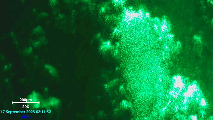
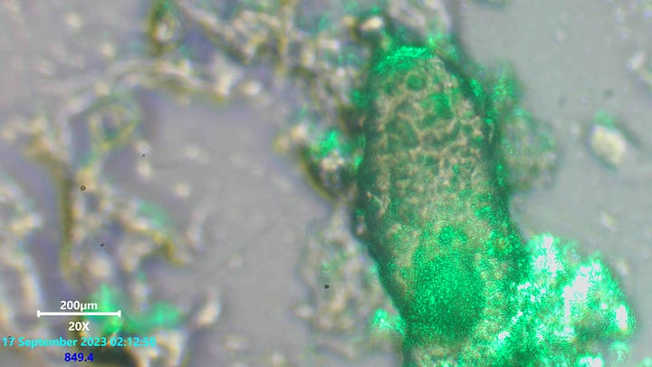
A mixture of compositions has been noted in the blood. The laser microscopy has revealed a repeat visual phenomena where curly interlocking borders of contrasted light can be seen on particular features. Again it is noted that just like when using polarized light techniques that reveal complex phenomena mostly with suspicious foreign artifacts that the same situation is true here using scanning laser microscopy.
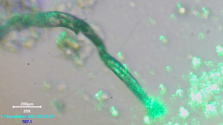
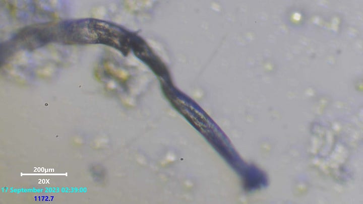
None of these patterns were observed with light or dark field microscopy. It was almost observed using phase contrast, this was much less clear or readable but certainly revealed same patterns on some structures. The fiber above manipulated the light in exactly the same way as some of the gels or affected blood cells. Not all fibers were coated with this material it seems and some gels of different texture, and visual appearance under light conditions did not show this same pattern under laser excitation.
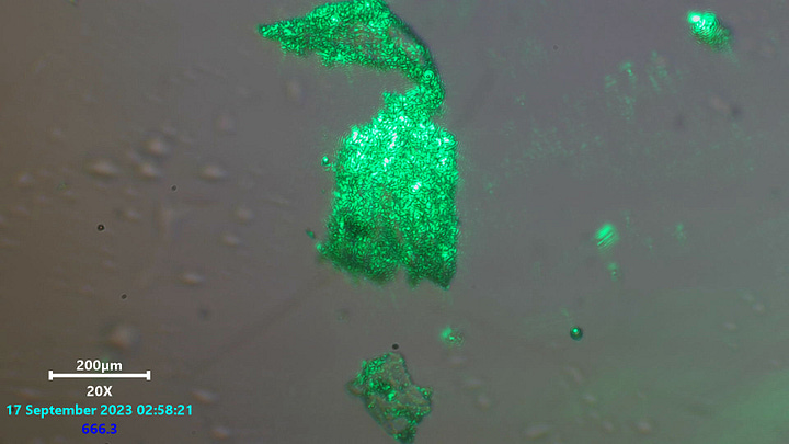
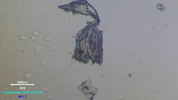
Amazingly even very different structures noted as being foreign to healthy blood also exhibited the curly squiggles though the structure via light microscopy seemed more uniform than gels and had many straight edge and stranded formations as seen in the image above to the right.
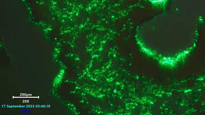
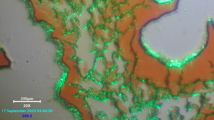
What has been suspected by many others also as not being cracks indeed have now shown behavior and criteria that does not agree with this once more commonly expected phenomena noted with normal blood drying and shrinking known in the past. Dr David Nixon among many has shown and seen these cracks to be filled with a gel like material often but not always containing what has been considered to be either Q-dots or CDB. Above we see signs of this same gel material interfering with the laser waves in a way that produces the same squiggly interconnected pattern as noted with the other foreign artifacts.
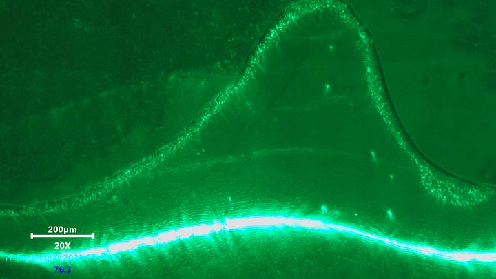
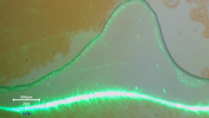
Blood bubble borders and streamed border lines throughout the blood show the same exhibition of this light phenomena too. much more than this has been observed using lasers on these bubbles too. Other structures, patterns and height maps have been revealed which highlight more than just blank space seen using dark field and light microscopy is really the case.
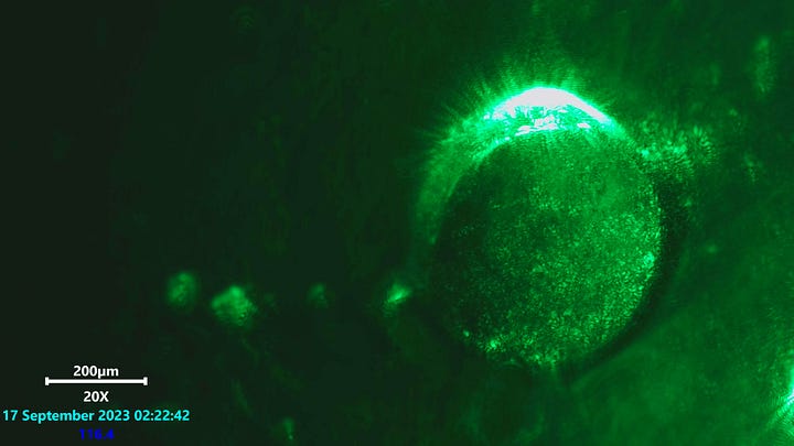
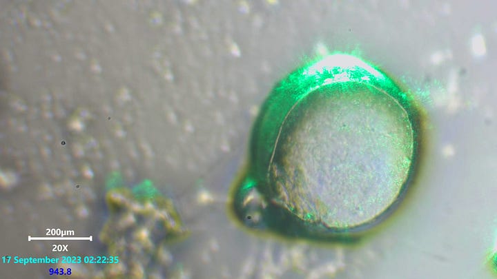
These famous spheres which have been spotted for many years and attributed as a familiarity known to morgellons disease have been observed crumpling, creasing, and going through various morphological stages not consistent with naturally occurring suggestions. I am still open here, but feel directed towards the likelihood of something closer to polymer spheres. The laser microscopy may be in fact showing these squiggles because of the possible polymer connection. More is being researched here and the discussion between others has been helping.
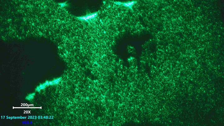
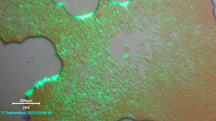
the above image shows the worse blood in the samples. As blood would group, smudge and show contrasted patches over time it would reveal the same curly excitation exhibited by laser microscopy.
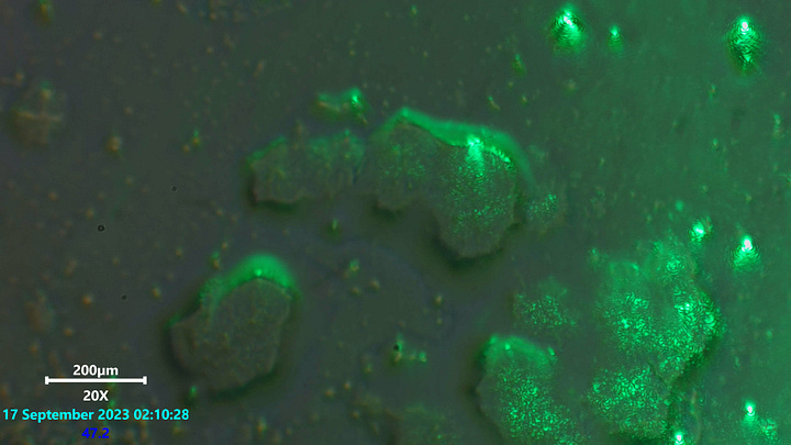
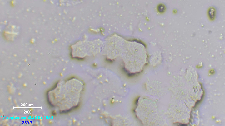
Some, not all structures of biofilm or gels would exhibit this exact same laser patterned phenomena.
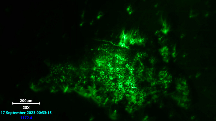
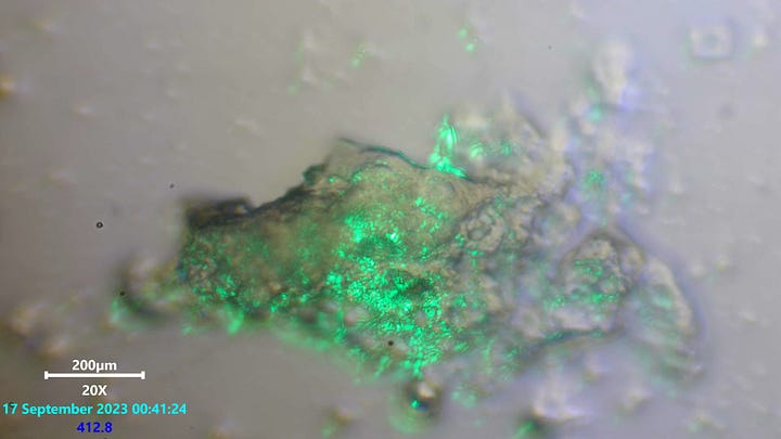
Another gel structure showing same phenomena. It should be noted that these were sometimes sitting on other masses of similar material containing differently opaque and colored consistency which did not exhibit the same light phenomena. A clear difference and a clear consistency correlation was made between the structures of question and the normal structures not unusual to the blood environment. More is being learnt from this end on what these interpretations may help in determining. Past matching of materials visually any other use of scanning laser microscopy is likely very limited. With the exception of detecting these same patterns moving erratically around the outside of blood cells at some points. A video of this shall be in the next post, it must be viewed on a large screen and focus must be applied to the details carefully. Either the material exhibiting this visual patterning is alive, or it is active by other none biological means alone. Please look out for the next post on this video.
Again i thank anyone supporting people who research these matters, not just myself. People are entitled and encouraged to learn freely in any way they deem fit, without intervention above the level of being disagreed with intellectually. My efforts are genuine and well intent. I leave myself open to education and debate by the traditional and honorable methods deemed humane to a free society. If i am proven to be wrong or having been misunderstood in my techniques of investigation i am willing to listen to others with knowledge in that field.


The Blob - glowing Neon Green
Hard to comprehend this was extracted from a human body
Amazing if unsettling work
Thank You
Creepy yet so fascinating! Thank you for the time and creativity- especially your openness to the scientific method playing out around the world now to try to unravel what the heck is going on and potential solutions. A gentleman you are of good caliber.