Bubbles in blood are possibly part of an in vivo Microfluidic system, an assembly system.
Unusual behavior of lipid structures in blood still under investigation.
We look into explaining the strange phenomena noticed while spending too long gazing through the microscope. A question a live blood analyst asked me not long ago “ what is with all these extra bubbles in the blood, they seem strange and there is more of them, are they bubbles? “
Before watching the live video let us consider that most of the recurring lipid structures we see occurring are mostly happening in connection with tubules and junctions, the yellow arrow indicates what i think may be a mixing vessel or tubule like the ones seen in Microfluidic processes.
Below is another example of video and tubules highlighted.
The above samples were taken from blood with added CoV swab solution to enhance and speed up the phenomena already seen now occurring in the blood for most. This work was done on a Leica DM2000 with industrial BIGEYE imager and using special 40x Oil immersion objectives. One of those things which increased significantly adding the swab solution was the activity of the bubble structures I have been previously studying in blood. The intensity and quantity of all the foreign material we are questioning so much in the last 4 years was far higher due to adding what I consider to be one of many significant sources of this product phenomena. Remember from previous posted videos that the swab itself under dark field looks almost identical to reactions seen in injectables by many the world over now, particularly by the first person to perform exemplary microscopy on the very complex crystal and dot structures it produces,
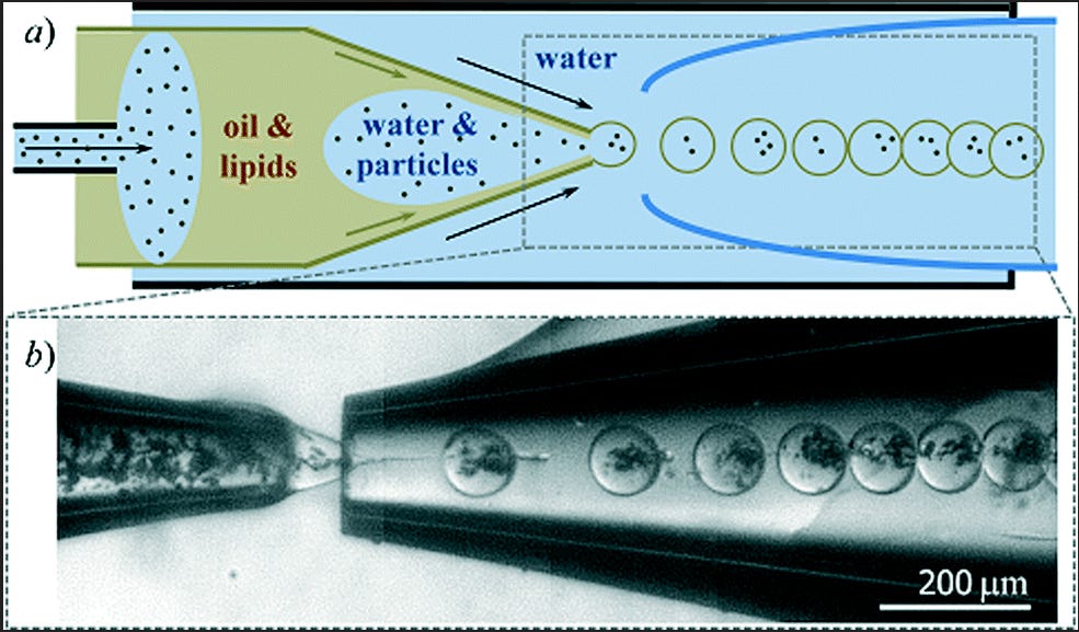
.The above image and website link shows a more simplistic form highlighting just one of the concepts possible using Microfluidic systems. They can become rather complex and evolve around the controlled feeding of specific materials through tubes which can form encapsulated packages. These capsules could explode rather instantly releasing nano sized or chemical payloads that may not be visible after, the can have time releases for drug delivery, or they can release via an external signal, trigger, or chemical event for example. The methods are endless, this technology has been perfected in vitro and in vivo, meaning self assembly of structures which can then form microfluidic systems for further self assembly, maybe even to fuel other processes which also may assemble more complex things from whatever was previously assembled at that stage. If a microcapsule is the desired product which i have also detected in samples via visual microscopy and detailed in previous posts then those capsules can allow proteins, DNA, nano-particles, and other stuff to be captured later if not at the time of encapsulation and that capsule then becomes another productive unit in itself.
Capturing these structures at the stage where tubules have formed and the activity is observed as in the above videos takes some time. The above videos of the lipid bubble action are still occurring now, almost 1.5 days later. We can tell the bubbles are not gas containing since the larger out membrane which seems to be the controlled environment for this smaller structures gets smaller over time and does not get bigger as you would expect if gases were forming inside of it. The above image shows structures not yet in configuration it would likely seem. Note the colorful blue dots for which i have found papers which might insight exactly what those are.


Enlarging the image on the left with light source lowered we can see various rings which look as though there are multiple layers of out wall. These varied slightly between structures and so did their colour. Some structures are perfectly round and we noticed that when they were forming they often did not form outwards in all directions equally. Sometimes the top what edge out, then the right, this very odd. Some of these structures formed, burst, and then reformed rapidly and others on different timed intervals. In one large cell there is almost a clocked pattern between all the forming structures and their release and this would make sense if they were producing something between them all and those things had to be of balance.


These smaller lipid structures contain a pattern of dots which repeats itself and can be seen forming without the encapsulation in earlier stages. The dots which are likely a mixture of structures and molecules are themselves able to create assembly of a stage when aligned in this particular way. It seems they can also do a great deal many other things too in other arrangements.
One will often not see the great colour, detail and same representation as i do without same or better equipment. It did not appear this way on my older scope and so people should note that while observing for themselves. In this image i show you that the largest amounts of the varying colour blue dots wish to be inside of a large membrane or what some might suspect to be an air bubble.
These layouts either had the outer membrane collapse, not form properly, or did need one if maybe it was supposed to construct other combinations not requiring the outer environment to be segregated. This also might not need an all encompassing membrane due to the fact it seems these bubbles actually seem to be the membranes themselves with the smaller forming lipids inside them. So these ones are different it would seem.


The configurations of lipid or cell’s formed in the sample does repeat its patterns often, but it can be daunting to follow with multiple variations repeating. This seems to be droplets forming on what looks like a long nano tubule feed line or a line used for spinning the formations we see here.The different light captures reveal the detail of the outer walls and the droplet towards the top right seems partly filled with a visible substance or materials.
There were many more observations taken in this long viewing, i will add those as separate modules in later posts. I will infer that what seems like fibrin spicules or spider web like proteins is actually seen so commonly now because i believe it is involved the processes we see happening. I believe that it involves calcium or calcium Ions and that this is why so many of us now suffer bone, tooth, and hair issues. I maintain the thought and belief that the added bonus chelating property of sodium citrate is removing this displaced calcium and interrupting processes causes by this nightmare. we need calcium for our bones and other functions but it seems that my hair has grown far faster, my teeth wearing far less and joints feeling much better for the multi attack approach that sodium citrate seems to be having.
(EDIT): there is a consideration in my mind that the expanding and contracting bubble activity could also be the result of PEG floating in between a phase where it is not able to go from one liquid state to another successfully.
As usual i spend every minute doing this stuff and never having any me time almost ever. Please help me to continue researching with others. I try not to post anything unless i see it as meaningful and useful to our global cause as free human beings. I get excited every time I get a genuine lead into identifying the processes here and even more so when i find something we can attack with natural products where possible.
I humbly thank you for your time and support. Thank you to those who help out with KO-FI and also to those who do not. buena salud para todos, bonne santé à tous, здоровья всем! (Good health to all in Spanish, french, russian.)


I believe there's 2 types of cold forming hydrogel we're dealing with. The Sodium Citrate deals with the calcium polymer. Thanks Karl C and Clifford. 👍☺️
The other cold formed soft white hydrogel, uses many Nano heavy metals as cross linkages, hence
the uniformity in its assembly. It also has a tunability, via photoswitchability.
The reason the polymers have acidic tendencies, the technology also uses a H+, proton pump.
The Amyloid fibrils are also part of the self assembly, it what makes the polymer hydrogels. Its process is similar to that of a Prion disease. The Amyloid elongate, become more polymer like and attach to the Nano heavy metals to form a cross linked mesh.
I'm going to write post soon. I got more literature to read, I think we'll have a protocol for disassembly and prevention.
Karl, You may want to check this medical science abstract-Health Effects of Exposure to Ultrasound and
Infrasound-pgs34-36. The bubbles may be inertia cavitation bubbles. If you check my blood, you will see thousands of micro air vapor bubbles. I am being tortured daily with sonic lasers causing acoustic cavitation of soft tissues especially abdomen where a lot of air and intestinal gas is accumulated causing stretching and enlargement of abdomen and internal organs and pain and suffering. When bubbles explode and collapse and during inertia cavitation they release mechanical pressure, heat, free radicals causing inflammatory, oxidative, hypertensive stress. Similar cavitation process occurs in boat propellers.
"2.3.3 Cavitation and gas-body effects
Acoustic cavitation may cause cellular responses as a result of mechanical, chemical or thermal means.
The inhomogeneous periodic field around a pulsating bubble can generate a small steady flow of fluid by a process known as microstreaming (Nyborg, 1998). The variation of this flow with distance from the bubble creates extremely high shear stresses near the bubble surface. It is now accepted that these
stresses are responsible for the observed temporary alteration in permeability (sonoporesis) and cell
membrane destruction resulting from exposure of cells in suspension in the presence of microbubbles. In
addition, mixing from microstreaming is the most likely explanation for enhanced drug transport through
the skin mediated by ultrasound. A bubble within a capillary bloodstream will stress that capillary when
forced to expand and contract in an ultrasonic field. These stresses may be sufficient to rupture the
capillary, allowing extravasation of the contents. Bubble collapse during inertial cavitation can also give
rise to a radiated acoustic shock wave into the surrounding medium. Inertial cavitation carries an
additional hazard for cells, free radical production. These highly reactive chemical species can be created
in the gas contents of an adiabatically collapsing bubble, because of the extremely high temperatures
and pressures created by the rapid bubble collapse. They may then be released to the surrounding
medium once the bubble fragments. However, in contrast to the association for ionising radiation
between free radical production and cell damage, free radicals produced by inertial cavitation lie outside,
rather than within, the cell. Whilst such free radicals may in principle migrate through the membrane and
damage intracellular components, both the presence of natural free radical scavengers and the very
short lifetimes mitigate against this. Were an intracellular bubble to undergo inertial cavitation, cell death
would be immediate. Finally, the presence of clouds of bubbles increases the absorption coefficient of ......"