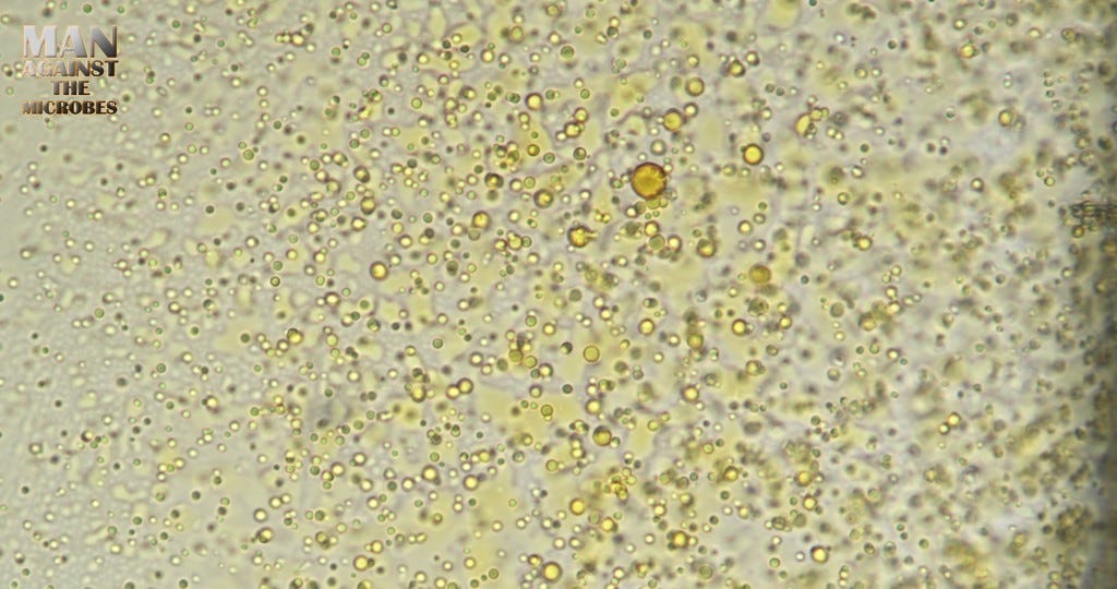
DYE techniques are proving to be very useful in analyzing COV swab (RAT) contents. Update
I have slowed down a bit since I have had to study into my observations much more deeply as we crack down on identifying the complex materials on swabs.
You wont find the answers here yet! We will see how far I have been getting with INDICATOR dye’s which do just that, They Indicate POSSIBLE CRITERIA for further analysis. I will summarize my findings soon since it takes a hell of a lot of work to get all these observations down, adjust the conditions, cross reference dye results, and research all the possibilities. Here are the raw notes straight out of my current experiments. I have been very excited by what i am seeing so far………. So here is stuff and things straight from my database notes !
Congo Red seems to highlight material in the Hydrogel matrix that is not apparent with Fast Green or Safranin.
The solution in general seems to not show coloration from any of these stains yet. Hydrogel may be inert and this is a known feature of PEG.
Some material that separates or forms does seem to stain with most dye’s. Fast Green occasionally shows what looks like it could be indicating Chitosan presence, but mostly shows more blue tones than it does Brilliant Green in some sample areas.
Not all tested drops from the same sample will produce the Brilliant green concentrations. Blue is always seen in large quantities, mostly towards the edge.
Lugol's iodine solution Will dye the early stage cells which look like bubbles.
Toluidine Blue shows possible indications of DNA and RNA.
Fast Green indicator on swab solution. Brilliant green is indicative of possible chitosan presence. Green is darkest where concentrated around and is also seen inside forming crystals.
Swab Fibers clearly showed uptake of the Fast Green indicator. Some turned blue instead, likely Because of a chemical interference of material in the matrix.
Blue was observed as explained before. Blue is highly unexpected using Fast Green and usually Only extreme PH difference and chemical reactions are likely to change the outcome to blue.
To see if blue resulted from PH I tried multiple samples with a solution of water mixed with Bicarbonate soda in order to raise the PH. The same blue phenomena occurred leaving blue as a likely result of a reaction due to a chemical present in the swab solution itself. At this time the Blue phenomena identity of the chemical causing it is of great interest.
Some of the formed polymer structures were able to show a green absorbance of the dye. The Structure is likely of mixed composition since it did not take the dye completely.
Turquoise to Green was observed using Fast Green, it absorbed into some fibers rapidly and began to flutter up and down the inside of the fiber rather unusually. Videos were taken.
These fibers were not formed but scraped off from the swab in to the sample. It is highly unusual for this behavior to occur in a simple textile like fiber of the sort expected to be used on a swab.
The addition of Congo red showed a vast amount of material that was not clearly visible Without this dye. Much of the material was of around 1-3um in size and would be seen in varying forms of distribution. The fibers which had FORMED in the sample were very compatible with the Congo red dye. It is of note that the swab fibers themselves seemed to absorb a very small amount but mainly showed externally attached materials which absorbed The dye far more and may have been a result of passing materials landing on the fiber.
Interestingly multiple dyes have indicate that not all the swab fibers are equal in composition. Some will respond to the dye very well, others will respond better to another dye instead.
Toluidine Blue dye exhibited colors that would usually be consistent with DNA and RNA. It is a dye that adheres to many acidic structures of other nature also. The indicator is as an indicator describes, only an indication of possible RNA and DNA which would not seem unlikely.
The same debris that are rich blue seem to also dye red with Congo red dye as above. Dots and granules in the background dyed mostly purple indicating possible RNA and Blue on the fiber indicating possible DNA presence. Not a solid analysis technique but good as an indicator !
The moving bubble structures which form at the edge of the swab solution droplet dye well using Lugol's Iodine solution. These seem to be capsules or synthetically produced cells
According to the observations I made using video of these sample types. The cells usually form contents inside and play an early part in the production of complex materials and chemical structures.
Before any conclusions are drawn as a direction for further indication I must do more staining.
Cross referencing all these multiple staining techniques will provide more confirmation of the likelihood that the dye or dye's may be inferring the right kind of material as strong suspects.
This will bring us to new levels of understanding and will help support or channel the direction of further analysis using other techniques such as HPLC(Liquid chromatography) going forward.
A few helpful but basic notes of interest from our database below. Too much on these topics to post.
If you are working with a PEG hydrogel or a PEG-based material and are looking to visualize or stain specific components, you may consider using dyes or stains that target those components. Here are a few examples:
Fluorescent Dyes: Various fluorescent dyes can be used to stain specific biomolecules within a PEG matrix. For example, DAPI or Hoechst can stain DNA, and fluorescein or rhodamine derivatives can be used to stain proteins or other cellular components.
Immunostaining: If you are working with PEG-based materials in a biological context, you may consider using immunostaining techniques. Antibodies labeled with fluorophores or enzyme substrates can help visualize specific proteins or antigens.
Ethidium Bromide: If you are working with PEG-based hydrogels containing nucleic acids, ethidium bromide or other nucleic acid stains can be used for visualization.
Congo Red: Congo Red is a dye that can interact with certain structural features in hydrogels. While not specific to PEG, it may be useful in certain applications.
It's important to select a stain based on the specific components you want to visualize within your PEG system. Always consider the compatibility of the stain with your experimental conditions and the potential impact on the properties of the PEG material. Additionally, consult relevant literature or protocols for staining procedures tailored to your specific application.
Congo Red is a diazo dye that has an affinity for certain types of biological and non-biological materials, particularly those with a high content of β-pleated sheet structures. Here are some materials that Congo Red is known to stain:
Amyloid Deposits: Congo Red is widely used in histology to stain amyloid deposits in tissues. Amyloidosis is a group of diseases characterized by the abnormal accumulation of amyloid proteins, and Congo Red staining can help identify these deposits under a microscope.
Cellulose: Congo Red can also stain cellulose, which is a major component of plant cell walls. This property makes it useful for studying plant tissues.
Certain Proteins: Congo Red has been used to stain certain proteins, particularly those with β-sheet-rich structures. It can be used in studies of protein aggregation and fibril formation.
Hydrogels and Polymers: Congo Red can interact with certain structural features in hydrogels and polymers. While it may not be specific to all types of polymers, it can be used to assess the structural characteristics of some materials.
When Congo Red binds to these materials, it exhibits a characteristic green birefringence under polarized light. This property is often used as a diagnostic feature for the presence of amyloid deposits.
Lugol's iodine solution is a staining reagent that is commonly used in microbiology, histology, and various other biological and laboratory applications. The primary use of Lugol's iodine is in the Gram staining procedure, a widely employed method for classifying bacteria based on their cell wall characteristics. Here are some common uses of Lugol's iodine in staining:
Gram Staining: Lugol's iodine is an important component of the Gram staining process. In Gram staining, bacterial cells are first treated with crystal violet, then Lugol's iodine is applied as a mordant. The mordant helps the crystal violet to better adhere to the bacterial cells. Following this, the cells are washed with alcohol or acetone, and a counterstain (usually safranin) is applied. This process differentiates bacteria into Gram-positive (retain crystal violet) and Gram-negative (lose crystal violet) based on the characteristics of their cell walls.
Staining for Starch: Lugol's iodine can be used to stain starch in biological specimens. It forms a complex with starch, producing a distinctive blue-black color. This staining property is often used in histology and plant biology.
Cytology: Lugol's iodine can be used to stain cellular structures in cytology, particularly in the study of cells and tissues. It may be applied in combination with other stains to enhance contrast and visibility under a microscope.
Observation of Protists: Lugol's iodine is used to fix and stain protists (single-celled eukaryotes, such as amoebas and paramecia) for microscopic observation. It helps highlight cellular structures and improves visibility.
Antiseptic Solution: Outside of staining applications, Lugol's iodine is also used as an antiseptic solution. It has been employed to treat minor wounds and cuts.
There will be alot more on this, but for now i just wanted to keep people informed of why i might be a bit slower to post lately. We want to get this right.
Thank you all for supporting, The materials and equipment are expensive. I manage all this research just on the odd donations so myself and others appreciate your help if you can. Use the Ko-FI button if you wish to support.


Thanks guys! Appreciate you all too!
Thank you so much Karl for your efforts. Frightening what has been discovered on the swabs.