FIBERS, SIDE BY SIDE, IN DIFFERING VIEWS.
Various microscopic comparisons of fibers for thought.

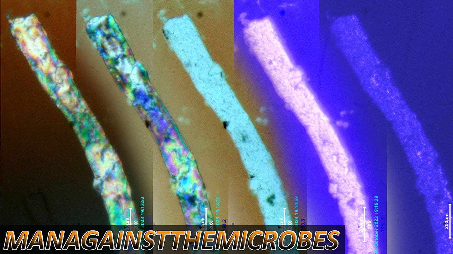
The two polarizing positions on the first two fibers reveal two actual possibilities of light wave by interpretation. Many internal details can be revealed with the likelihood that colors represent some differing material composition or structural organization inside. Bright field with increased contrast reveals mostly the dots and pits within the structure. Centered UV exposure reveals surface textures and what seems to be square crystal like structures embedded in the fiber structure. The indirect UV shows us less to some degree than the bright field image. The square crystals inside the fiber were not really noticed in untouched bright field. These were interesting features i have not noticed before.
The textures noted using polarized microscopy are not easily or not at all observed using bright and dark field microscopy. These details are useful when comparing these fibers to textiles and Nanofibrils in scientific literature.
Pits and what some of us think might be Q-dots or CDB can be seen clearly in bright field with increased contrast editing.
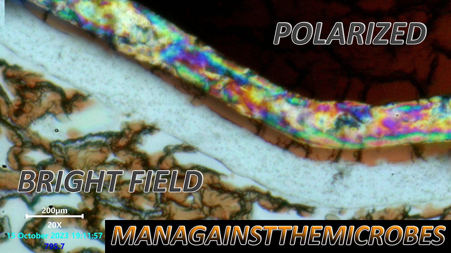
Above image details a strange fungal looking mass wrapped around the fiber like a bandage. Bulges and other 3-Dimensional geometry can be seen using polarized filters that is also not visible or observable in bright field. Seeing the light waves in the polarized form allows us to see much detail which cannot be perceived using other techniques.
I shall continue using adjusted forms of polarized light microscopy and see how far its use can be applied. For now it is an interesting method still and offers detail to examine from alternate perspectives.
I am using KO-FI right now and appreciate you clicking the link to go there and contribute.
Thank You All !!!!!!


This is amazing work! Thank you!!! ❤️
GREAT WORK VERY MUCH APPRECIATED: THE MULTI COLORED FILAMENTS ARE PRE STAGE PACKED CHEMICAL PROCESS NANO PATHOGEN PRINTER CHIP COMPONENTS PREPARING FOR POST DISCHARGE SUB STRAIGHT PC BOARD LAYERING & ASSEMBLY THIS WAS FIGURED OUT IN 2012 WHILE SCOPING CHEM-TRAIL FILAMENT GENERATING NANO UNDER SKIN FUNGUS OUT