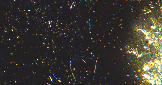
HUGE! Photonic dot activity captured in slow motion using an agent to slow the technology down!
Our sample is just tap water with an agent added to slow down the action.
Lets go down to the disco!……. Oh no, that is my tap water on a slide.
I have informed a whole bunch of fun people on the research chain that we can now slow down the technology in samples to see what we would not be able to catch normally. It is almost too much to bare knowing i just bathed in this. The water in my bath now runs green! Check yours, remember when it was almost clear or blue. Well that has to do with the tech having a green tint when viewing with bright field microscopy. Now your utility water does it too.
I am not sure many will believe there eyes here. David Nixon was watching my scope live when this happened. I got all loud and crazy when i saw it.
The video above uses our slowing agent which allows the dot activity to be seen much slower towards the drying of the sample. The material absorbs water rapidly, but we used something that gets it wet and active but without the material being able to absorb that liquid so rapidly. The dots can be seen pulsing, flashing, moving, and aligning. They do this in order to form structures in so many different ways that yet again that part is unclear as to what they are forming other than crystals, gel artifacts, fibers, and much more
You can see the dot structures above forming around the circular structure and then filling in the space around, If you watch via the big screen it is a whole different experience watching each of the dots move, align, and light.
Above is a less obvious display of slowly changing bright dot streams changing behaviors. Shame they were not going to any music since that would make them even smarter. The next video below is a real killer. We can see the drop of tap water without the agent used below. A few dots are seen floating around in our drop of tap water at first, and then we them slowly getting larger, more in quantity, and then forming together as larger structures. The process can be repeated over and over.
Here is a run around the slide, we can see all the stuff found in blood samples if we go by a visual approach. Doesn’t take a genius to see the relevance here.
So the case in hand shows that looking through my previous observations that there are biological elements implicated and also nanotechnology ones which we clearly see in the videos above. The transhuman package is coming together and is a very disturbing reality for all.
I need to catch some sleep since i have been on the scope far too long everyday on top of all the great research talks. The community has made some good suggestions along the way and you have all been a really good bunch of participants so far.
Hopefully all the other researchers around the globe who have been shown this phenomena elsewhere before posting can now realize the need to decipher the chaos more critically. Seeing the implications of all the findings here on this stack and on others should be encouraging to those who know more in this field, people need you. I cant wait to see what everyone else discovers about what we are seeing from my microscopy, research and others research/microscopy work.
Please don’t forget to help out if you can by supporting us with KO-FI, Thanks!


Karl, I wrote you (it seems) in Telegram.
I’m ready to give away electron microscope for your research or for one of your trusted partners, please let me know either here or in telegram.
It’s in Berlin currently, not really sure it’s possible to send it to USA, but probably you have interested colleagues in Europe
I've just looked at our filtered (Carbon 0.2micron) rainwater we use for drinking that we collect from a property about 400Km north of Sydney (NW of Kempsey) & found nothing in it so then checked water from the tap at our living space on the Central Coast NSW & found a large cloud of microdot things in that. Unfortunately my pathological microscope doesn't have a camera attachment but does have a dark field filter which allows me to see this junk material. I only have some old slides that appear to have micro scratches & what looks like mould spores that I can't seem to rub off even with something like windex. I'm curious to know Matt whether you have checked bare slides to see if there is any pre contamination before adding specimens since a lot of what I've seen on mine does look a lot like the things I am seeing in yours & David Nixon's images. I also note that after making some colloidal Gold & filtering sediment from the new batches I get a collection of what looks like coagulated Gold micro clusters in the filter paper. This stuff seems to occur more often if I've left my electrical equipment on & the spark gap has collapsed leading to a direct heating effect which then precipitates the Gold nano particles out into a black sludge. I've then looked at a small lump of this stuff under a 40x scope & see these tiny fibers coming out of the sample that also remind me of those I'm seeing in your slides.