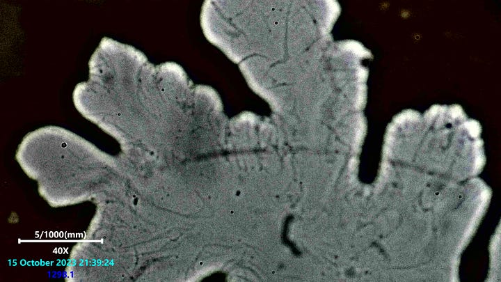
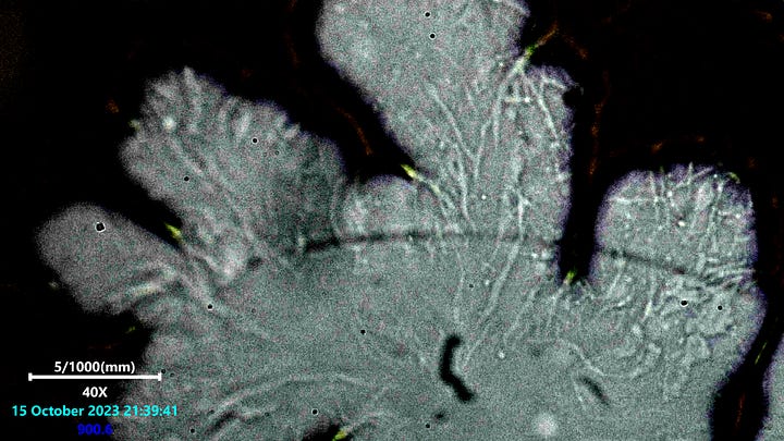
More evidence for large bubble voids in blood actually being full of material.
Multi-layer imaging of blood bubble structures.


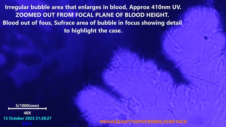
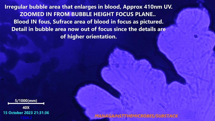
It has become clear that 410nm light wave (UV) does not always reveal the presence of material in these void areas. Attempts on some of these structures using UV revealed very little this time around. However nearly every bubble structure contained these snotty looking traces that one would assume are either bacteria, fungus, proteins, or possibly even something else.
These structures are being narrowed down by process of elimination and cross referencing criteria obtained by several methods used in my research. Those methods being polarized light microscopy, laser scanning microscopy, dark/bright field microscopy, staining, and morphological features. This is a good start but will still only serve as a pointer for further analysis.


UV had a feature not included with bright field which supported differently the fact that these structures contain material otherwise previously thought to be empty bubbles. on the left zoomed out we can see the thin line looking details from structures inside the bubble overlap the surface of the blood. That phenomena was not seen using bright field microscopy. It is also not seen when focusing in deeper to the slide level as pictured right.
Many methods have been used already to confirm that these voids are not empty space, but clearly these are more likely to be a more complex structure. Either these are part synthetic structures or they are organism based structures which might even be seen as a whole animal. The fact these grow, detach, and behave in no particular pattern might imply a unicellular organism like slime mold. One can only have ideas for now, just thinking out aloud and sharing those thoughts.
Up till now the evidence for these not being bubbles from my own analysis are as follows:
Note that i have considered that water, fats, human lipids may be separating from the blood plasma, or from the crumbling RBC’S. If this were the case i would think to expect that these things would separate in many areas and not just from locations originally seen at the time of slide prep. It is literally as if these liquids only for from the original sites and grow outwards.
The vein like snot features can be observed in most all bubble like structures using various techniques. Not always visible using bright or dark field !
These vein like structures have been observed moving in the liquid or gel like mass of the bubble using time lapse. They move slowly like cobwebs in wind. It is very hard to notice without very high magnification and a keen eye, but indeed are moving.
Under bright field green lipid domes have been seen in many of the more circular bubble voids. They are also seen moving and likely implies they are in a wet material or gel mass.
Laser scanning microscopy has shown there to be detailed height mapping to these structures as seen in previous posts.
Material such as crystals and ribbons form within these structures often which also would imply that enough surrounding material must be adjoined or in contact with such grown structures.
UV microscopy at around 410nm has occasionally revealed cloudy masses in some of the bubbles, this could not be seen with other microscopy methods used so far.
The larger bubble like structures will grow and eventually take over large areas of the slide after a few days. Something in these structures must be consuming substance of some kind in order to increase in mass.
It is known that what we see here is part of a phenomena known to the medical industry for many years. Information on this has seemed lacking or as if missing part of the picture. Is this something that has been with us for 45 years or a 100+ ? It has become reasonable to question any occurrence noted within the blood given the situation we now find ourselves in. Something may have been altered, manipulated, or be miss understood which has great significance to the problems we face now. Answers are often found where we least expect them, it has been noticed that fibers and other ribbon like structures have been seen forming in these areas of the blood. There seems to be something going on here, in plasma, and inside RBC’s. I sometimes wonder what Clifford Carnicom has observed of these bubble voids.
If anyone has seen any previous peer reviewed journals on this topic then please do share with me.
New lab gear is needed ;D......Thank You All !!!!!!


Love it! I know something’s up w these “bubbles” or isolation zones..........
Nice work Karl, thanks. Give me a month to get on top of some bills mate