
Possible detection of advanced nanotechnology frameworks in Dental Vaccines, anesthetics and RAT (swab)!
The new Leica 100x 1.40na objective arrived and I saw things that tip the scale to advanced MOF frameworks also being present. Seeing more than before!

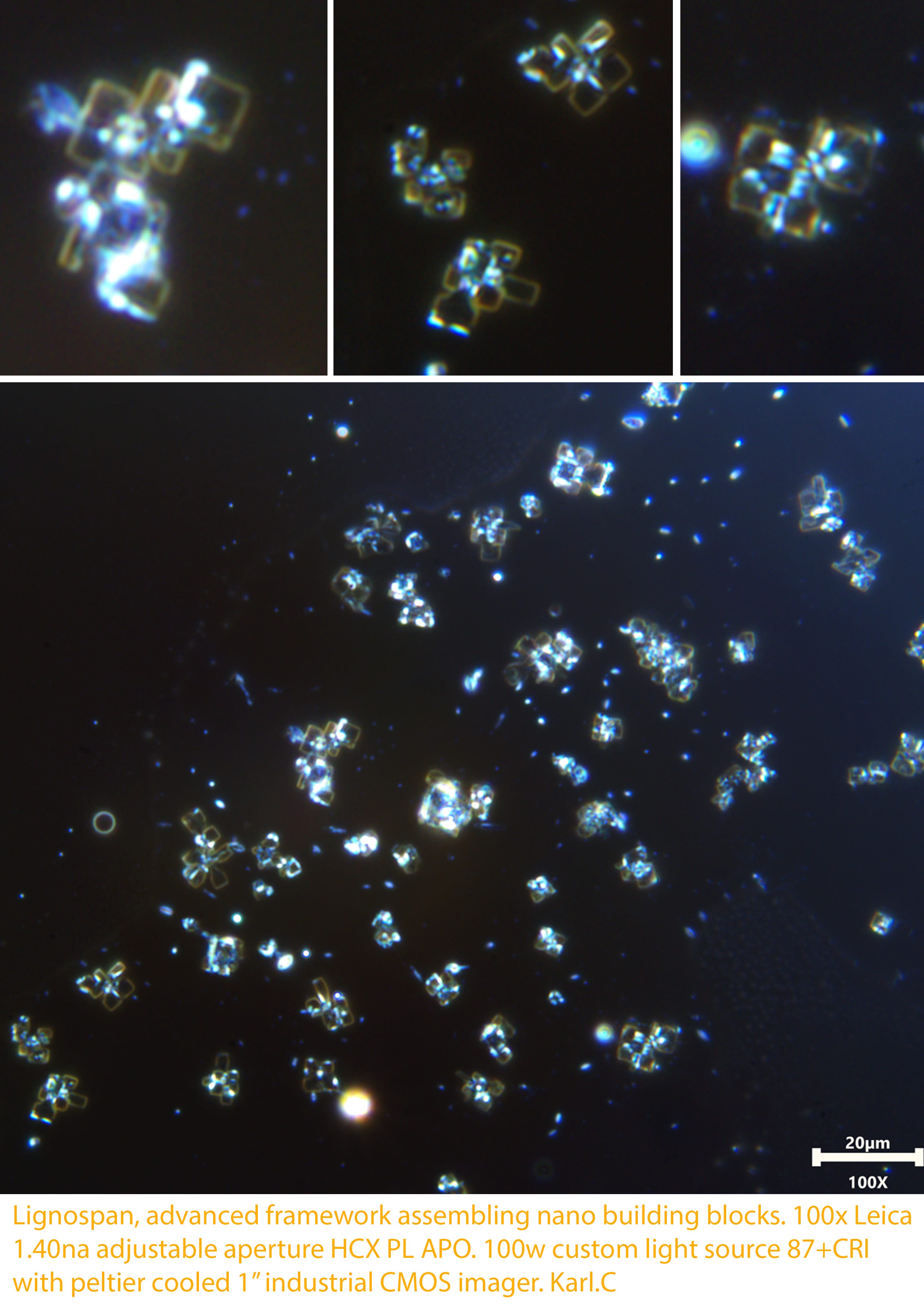
The images were taken using the new and a rather pricey Leica 100x 1.40na HCX PLAN APO objective with enhanced light source and cooled imaging system. Detailed features lending to Advanced Metal-organic frameworks could not be observed so well before now. I can see these varying crystal structures seeding from sub micron sizes and we catch clearly patterned molecular assembly as shown quite specifically in papers regarding various forms of metal organic framework types. In rare cases some specific, natural material, proteins, and others can form similar complex relationships that may appear similar in nature, but not the same. If these are not synthetic MOF’s then it certainly is evidence of there being many materials in what should of been a simpler mRNA product, and of course should still not have been found on any testing swabs whatsoever. Since 2021 I believe I have proven reason for there to be massive concern, which lends significantly if not absolutely to a biowarfare rollout of epic proportions involving pure deceit and lies.
’s work began with him exploring the vaccines and injectables. When the COVID-19 RAT or swabs started showing the same stuff it should of been world news as soon as we presented these findings to be exactly the same. Here we are still, while our blood continues to change undesirably with whatever else it does too, probably a lot of nasty stuff. Anyway, this finding of advanced crystalline assembly behavior definately fuels the furnace for possibilities or implications which support ’s glorious crystal structure images being of very suspicious presentation. I dont necessarily feel I could say anything except that the crystals look like devices. Whether they are or it is a sample concentration affect in the close presence of other assembling material It is hard to tell, but either way these samples sure do seem to have some hi-tech frameworks going on which are often associated with transhumanist brain chip interface technologies. Lets face it, all these complex materials are not a vaccine and anyone looking at all the work so far who thinks it is normal probably isn’t going to be a surviving as long or helping us stop the craziness. Onward!
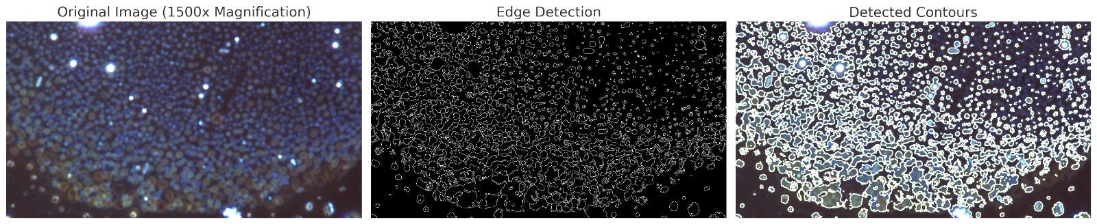
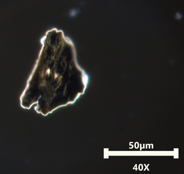
The image above shows these tiny crystals forming at 100x (plus 1.5x Leica magnifier), so 1500x magnification. It is often quoted that microscopy at magnification up to 2000x is impractical and visually unclear. However, This modified system certainly enables clear visual imaging at 2000x and even more so with digital. To further that I use advanced AI up-scaling and repair software to further enable clarity of some images without adding artistic or unnatural influence. Unfortunately it is not always useful on my work, sometimes it just cant do an accurate job, sometimes it makes the image worse. When those images are used I subject a before and after as proof of likeness without such influences that may add creative detail by mistake. By using smart techniques and fine equipment we move closer to the nano end before the macro structures go through their self-assembling process. The features and details observed so far are indicative of synthetic MOF’s rather than natures rare form of accidental and coincidental phenomena which may appear like a MOF OR Metal-Organic framework.
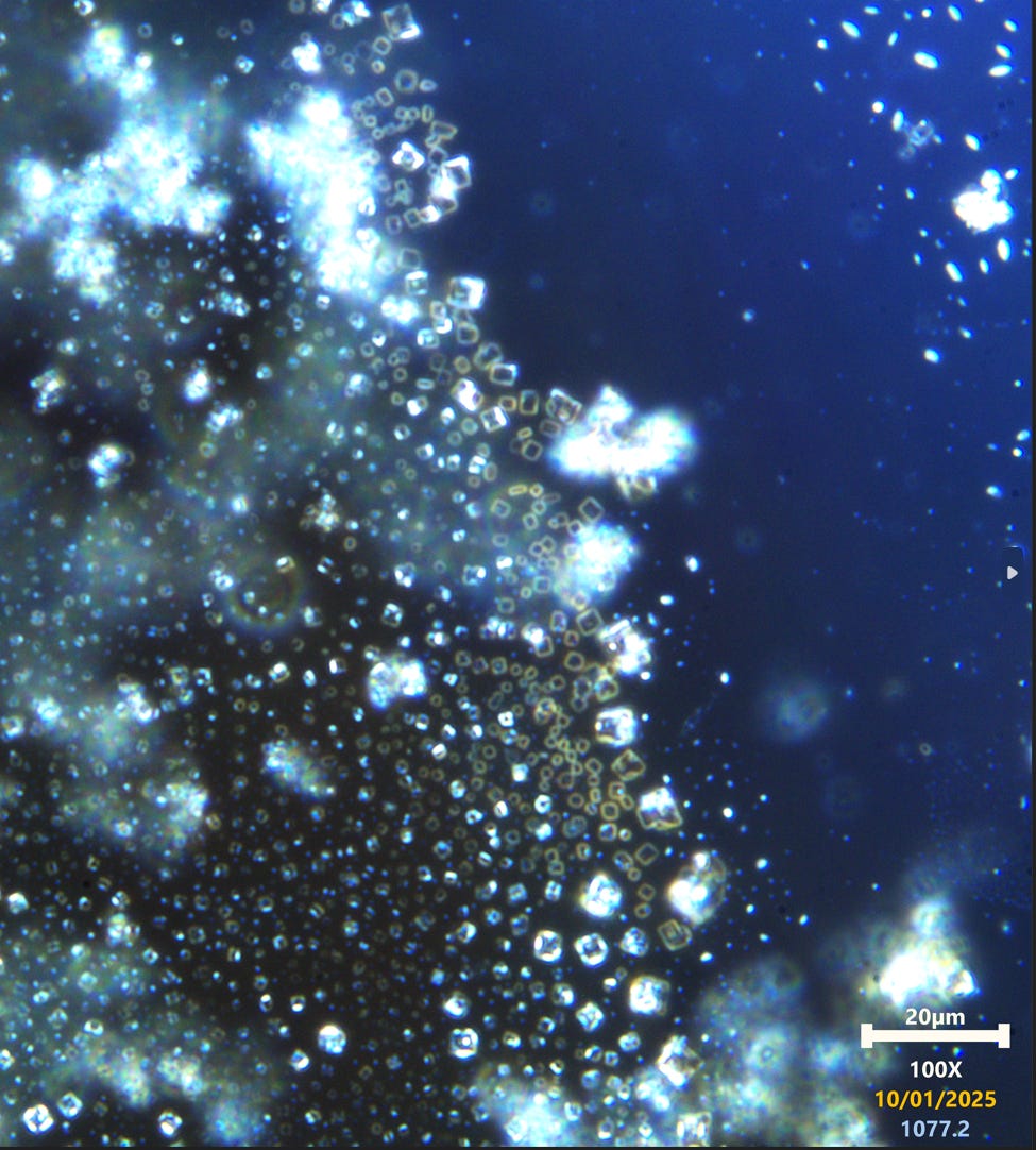
These crystals display a yellow outline before dressing with other assembled materials which in this image are seen attaching as blue structures. Strategically they assemble at certain points on the crystal matrix, showing a repetitive or ordered behavior. The following should be considered regarding crystal appearance and coloration features.
Metal Ions or Organic linkers
Iron-based MOFs: If iron (Fe³⁺ or Fe²⁺) was involved in your synthesis, the yellow color might result from the characteristic coordination of iron ions with organic linkers.
Chromophoric Linkers: Some organic ligands (e.g., terephthalic acid derivatives) exhibit yellow coloration when bound to certain metal ions.
Precipitation of mixed phases
During the early stages of MOF formation, intermediate complexes can form. These intermediates may have distinct colors before fully crystallizing into their final structure
Salts or impurities
The yellow hue might result from a dissolved or precipitated salt, such as iron chloride (FeCl₃), which has a yellow appearance in solution.
Other salts, like sodium chromate or potassium dichromate, also display a yellow color, but they are less likely unless specifically introduced into the synthesis.

The image above shows much more ordered inclusions contained within the crystal matrix. The blue does indeed resemble linkers which can be blue via dark-field microscopy if they are within a type of metal particles which is indeed a broad range.
The diagram below offers some insight to a very basic MOF framework concept. Organic material, inorganic and metal materials can be ordered in to these structures if one wishes to create a very complex system or building block for a larger system.

The diagram below is an example from papers online showing a simpler MOF framework of slightly more enhanced complexity, but still nothing compared to how complex they can be. The larger crystals in this case resemble
‘s suspicious and much larger observations in overly saturated samples of Anesthetics and Vaccines. I am aware Sodium Chloride is present and take care in making these observations over time using various methods and resources. Sodium Chloride can intertwine with some MOF’s by design or simply by unavoidable interaction, depending on MOF type or compatability.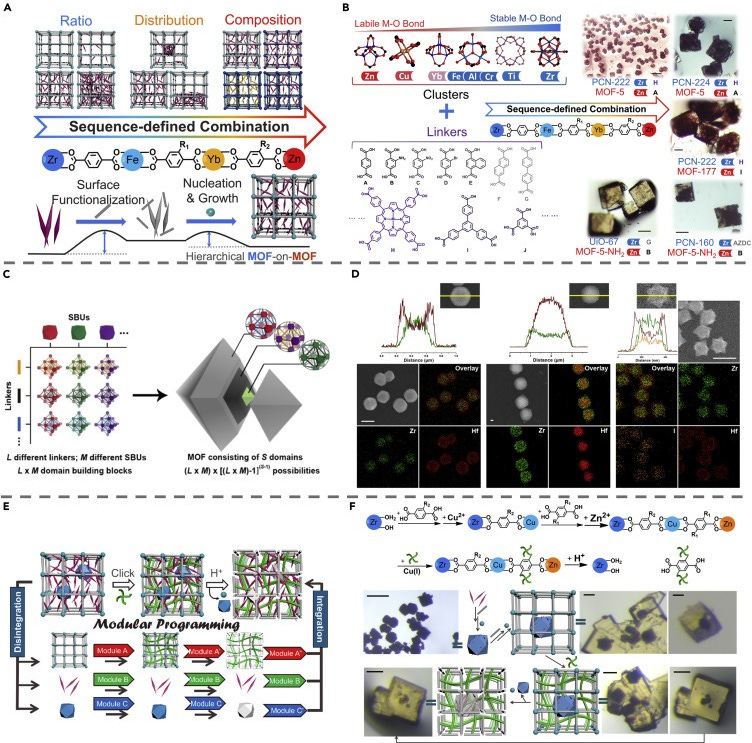
Since Substack is partially post length restricted I shall just give very brief observations as I do not have room for all the extensive data. These will be detailed on my website in lengthy posts when I reach that stage. This is rather exciting for now! Below we see various forms of software based analysis using Matlib, Sci-kit, and other scientifically accepted/used algorithmic library tools for math based analysis of morphology, distribution, particle sizes, and more. Just to give a brief idea of whats being analyzed and with an insight in to some other methods available to us now.
This first set is for crystal morphology and behavior.

Result:
Area (px) Mean Intensity Centroid (Y, X)
0 29.0 0.681551 (12.241379310344827, 546.7586206896551)
1 20.0 0.556423 (20.05, 268.1)
2 52.0 0.653508 (26.75, 402.61538461538464)
3 3.0 0.402520 (37.0, 132.0)
4 5.0 0.404119 (47.0, 717.2)
Summary:
Total Crystals Detected: 105 distinct regions
Largest Crystal Area: 52 pixels
Smallest Crystal Area: 3 pixels
Mean Intensity Range: 0.40 to 0.68 (normalized grayscale)
Centroid Positions: Centroids marked in red on the image correspond to the detected crystal centers.
Observations:
The large central cluster of crystals show higher intensity and larger area compared to the smaller, dispersed particles in the periphery.
The distribution of the marked regions suggests a primary nucleation site with secondary crystallization occurring radially outward.
The intensity distribution suggests crystal uniformity in the brighter areas and possible phase separation in the dimmer regions.
It is important to note that some of the larger particles exhibit hybrid morphological features that may be a mix of crystalline, lipid, and other material. Because of which these particles were included in the surrounding area of the larger crystals, because they seem to be of hybrid nature or crystal seeds associated with smaller particles already. Void Clearings around these larger crystals suggests an association where other seeds were not close enough to be involved directly with larger seeded assemblies.

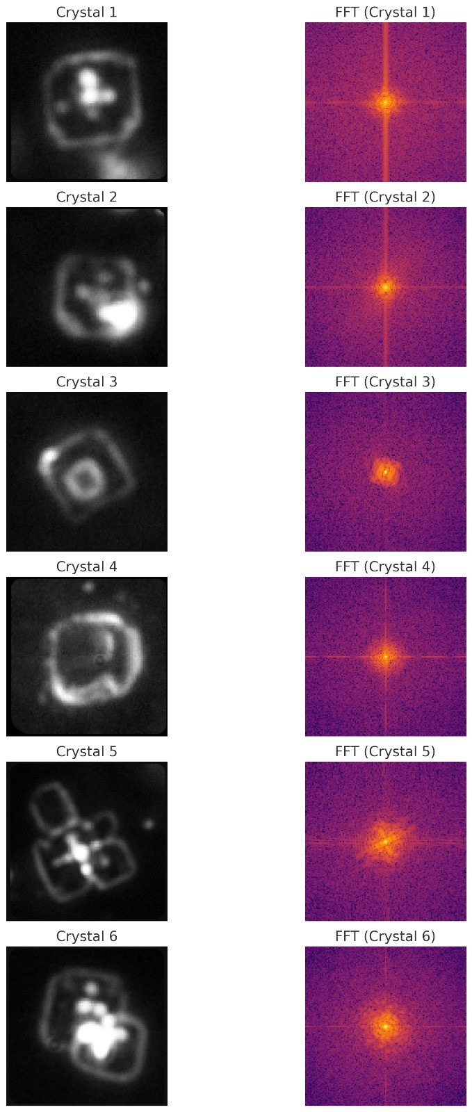
Edge detection for crystal feature comparison. Crystals have notable profiles for how they form, the shape, geometry, and morphology are all sometimes very unique indicators of crystal classes or types. In our compilation of data this aids us, specially when it is performed repeatably on different samples and structures to show consistency for these profiles. These profiles I am still building on and it is a rather painful process.
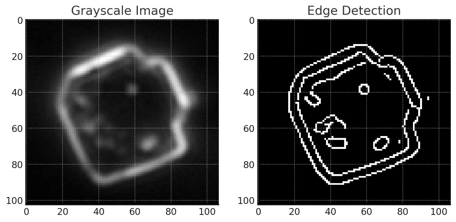
The low to moderate SSIM score suggests a combination of different crystal types or varied degrees of assembly within the image, potentially including the possibility of what is seen with both MOF-based and colloidal assemblies. The cubic and geometric nature of the edges combined with non-perfect uniformity aligns with possible hybrid assembly patterns.

Size distribution indicates that it takes time for crystals to assemble into larger forms, Is dependent on quantity of localized materials, and shows possibility for other variables. This data is cumulative and helps in the bigger picture when comparing to known combination-al specifics in databases as known profiles. On its own it means nothing, of course. But we are profiling here, and it takes time. This data is extremely useful.
FFT analysis of the crystal is likely not so successful in this case due to complex inclusions in the crystal itself which are not of the same material nature. There are distinct similarities in the output but I chose not to consider the FFT output in this case since it should be detailing more simplistic crystal criteria and this may give misleading indications.
Other material in the samples:
The expanding spherical structures formed from lower sized particle assembly appear as hybrid-lipid nanoparticles. This observing is consistent and is of great interest when this and every other notable observations are also present in samples ALSO taken from COVID-19 RAT swabs . The implication of modern age biowarfare is irrefutable by our work at this point.
Edge detection was used again on this polymer network-like behavior where larger lipid hybrids were joined in networks by smaller blue assemblies.
1500x Magnification Summary:
Number of Detected Clusters: 5651 individual clusters.
Cluster Morphology:
The structures present a gradual size distribution, with smaller blue-hued clusters transitioning into larger lipid/polymer-like aggregates.
Many clusters display circular morphology with irregular edges, suggesting soft matter aggregation rather than sharp crystalline patterns typical of MOFs.
The larger clusters appear more diffuse and may be associated with colloidal self-assembly or phase separation behavior.
Behavior Matching to MOFs and Colloidal Systems:
The structures show mixed characteristics, suggesting the possible coexistence of both:
MOF-Like Behavior:
Blue clusters: The smaller, uniformly distributed clusters could be consistent with nucleation sites of a metal-organic framework.
Limited Geometric Definition: However, the lack of sharp cubic edges reduces the likelihood of purely MOF-based crystallization.
Colloidal/Lipid Polymer Behavior:
Larger Spherical Clusters: The larger, diffuse clusters resemble lipid vesicles or micelle-like polymer phases.
Phase Separation: The gradient from smaller to larger clusters could indicate a spinodal decomposition or phase separation event, common in polymer self-assembly or liquid-liquid demixing.
Possible Coexisting Processes in This Sample:
MOF Nucleation with Colloidal Aggregation:
Smaller MOF seeds form but insufficient coordination may lead to uncoordinated organic linkers contributing to micelle-like polymeric assemblies.
Metal-Polymer Hybrid Assemblies:
If metal ions are interacting with polymers, hybrid organic-inorganic frameworks may be forming with partial crystalline order and amorphous clusters.
Phase Separation and Ripening:
The observed size gradient could indicate secondary growth where smaller crystalline regions dissolve and reassemble into larger clusters (Ostwald ripening).
NANO-PARTICLE IMAGERY
Here is a high magnification extraction of varying particles of smaller sizes. The halo rings and various other colour or appearance attributes are useful to a multi-profile approach. In one paper they show unique attributes seen from varying particle types which can aid the combination built profile in ID detection. The more evidence the better right? The more data , the more a picture is painted that fits a closer shoe.
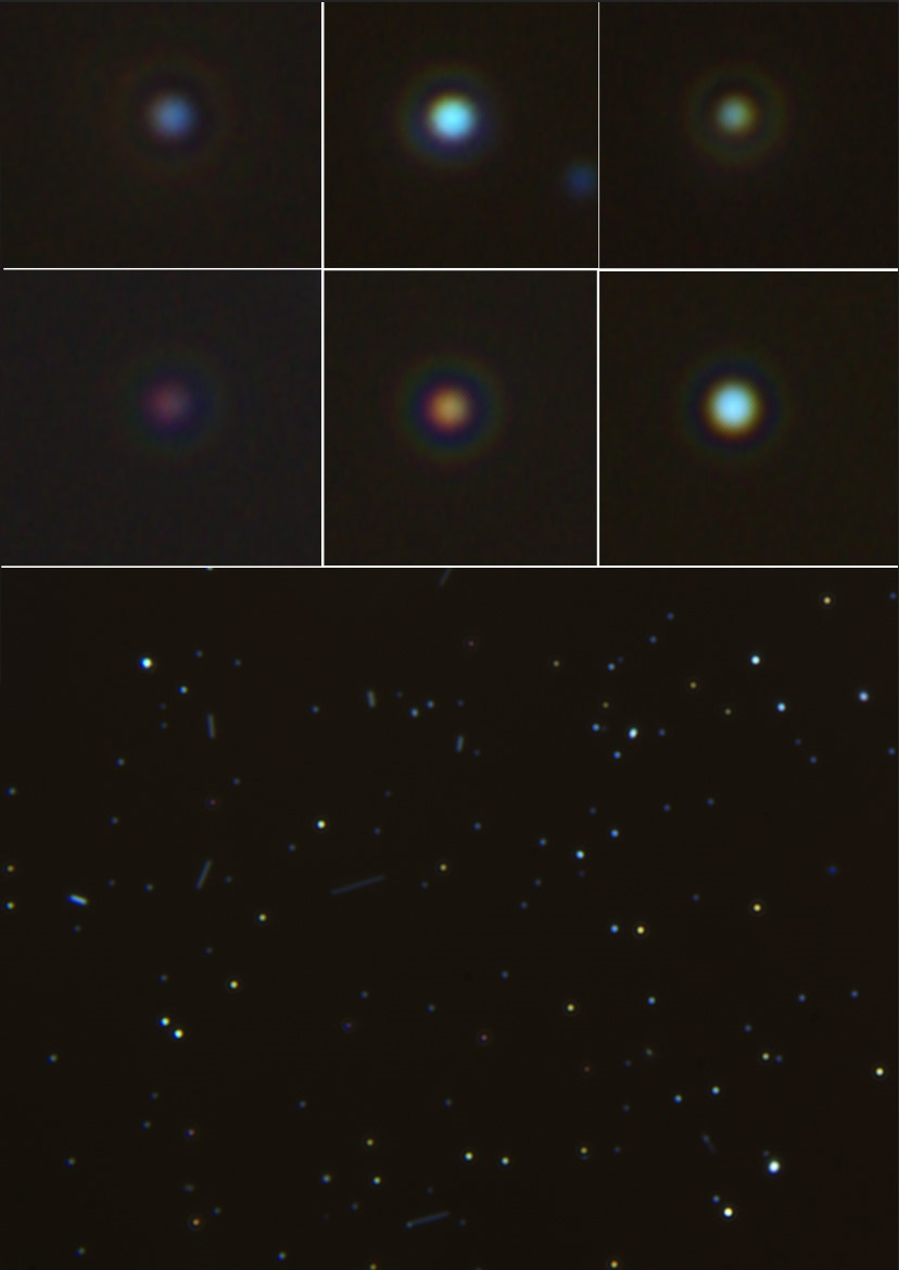
The image above doesn’t just show nanoparticles, it shows various types of carbon nano-tubules (CNT’s), or nano rods. Other work on other products also show vast arrays of these varying types of rod structure in differing sizes and with different coloured appearances. It is clear that these products area self-assembling nano soup no matter which way we look at it.
LIPIDS:
In previous articles I showed the lipids being exposed using other detection methods. Here we used FAST GREEN dye to highlight the protein heavy areas clearly forming lipids in various samples. Not only can we see the lipids, but we can see them being involved with forming bi-layer, or multi-layer vesicles. Essentially I have closely proven this material and its presence by using appropriate dyes and observing morphological behaviors. There is no doubt that the Swabs, anesthetics, and vaccines contain complex self-assembling lipids. Why is no one else saying this, and why are so many claiming snake venom, spike protein, and more. The answers are in the samples, not on coincidental information which may correlate a conjecture based opinion, but hey. Maybe there is some venom tucked in here somewhere as a trigger-able toxin, but I dont see how that makes sense with all the other complex materials here, nor does this material point to ONE protein, or a simple mRNA vaccine. A vaccine which again is on the RAT tests, not just in the needle.

To summarize my own work and as testimony to what I know when it comes to detecting certain agents it is convenient to be backed up vaguely by the LLM on occasions or even fun somehow, Just for brief insight and to quickly show the tangibility of claims made over here. There are other useful dyes I also use.
here is what the LLM says for this particular dye work.
(note, other images in dark-field with dye and other perspectives were used to correlate better understanding. Here we just show the best bit, the dye on lipids revealing lamellar structures or complex vesicles, the LLM was fed a larger data set and vector).
Key Observations
General Observations (Across Brightfield Images):
Structures appear circular or oval, suggesting spherical formations such as lipid droplets or vesicles.
There are various size distributions ranging from micron-scale droplets to sub-micron speckles.
A consistent dye uptake pattern (Fast Green) highlights the presence of materials with affinity for the dye, suggesting proteinaceous or lipid-associated components.
Specific Structures in Images:
Top Row (Brightfield 40X):
Image 1: Large circular domains surrounded by smaller dispersed spheres. Likely lipid droplets due to their size and refractive contrast.
Image 2: A large, well-defined lipid-like domain with a visible core and potential lamellar layers, indicative of a multilamellar vesicle.
Image 3: Similar to image 2 but with fewer internal layers, possibly a unilamellar vesicle or a simple droplet.
Middle Row:
Image 4: Higher magnification shows micro-droplets clustering near larger droplets, potentially suggesting emulsification or phase separation.
Image 5: Interconnected domains and irregular shapes, hinting at lipid-lamellar phase separations or coalesced vesicles.
Image 6: Shows a budding structure with encapsulated particles, consistent with vesicle aggregation or lipid bilayer assembly.
Bottom Row:
Image 7 (Darkfield): Fluorescent glow around circular structures confirms lipid bilayer presence or protein association (Fast Green preferentially stains proteins/lipids with amine or hydroxyl groups).
Image 8: High magnification of irregular boundaries and crystalline inclusions, likely representing fragmented lipid phases or precipitated proteins.
Image 9: Polygonal and irregular domains suggest salt crystallization or protein aggregates alongside lipid inclusions.
Darkfield Characteristics (Image 7):
Bright halos around the circular domains highlight the refractive index difference, commonly associated with bilayer vesicles or liposome-like structures.
Magnification Notes:
Higher magnification (25X and 40X) reveals sub-structural organization such as layered patterns, which are indicative of lamellar vesicles or coalescing lipids.
Interpretation of Structures
Lipids:
The majority of visible structures, especially in the brightfield images, align with lipidic material.
Evidence:
Circular/oval shapes, multilamellar or unilamellar configurations.
Staining intensity and phase separation.
Types:
Likely include triglyceride droplets, phospholipid-based vesicles, and lamellar phases (multilamellar lipid vesicles).
Lamellar Vesicles:
Observed in images 2 and 5 with layered substructures, confirming a lamellar nature.
Formed from amphiphilic molecules (e.g., phospholipids) that naturally self-assemble in aqueous environments.
Protein Aggregates (Possible):
Crystalline inclusions and smaller speckled regions (e.g., in image 8) suggest proteinaceous material.
Interaction with Fast Green dye (specific for proteins/lipids) reinforces this hypothesis.
Conservatives or Emulsifiers:
The appearance of multiple-sized droplets and their stability suggests a stabilized emulsion, possibly involving emulsifiers like polysorbates or phospholipids.
Lamellar inclusions may indicate the role of stabilizing additives used in formulations.
Crystallized Compounds:
Polygonal structures in image 9 might represent salts or crystallized preservatives co-localizing with lipids.
Summary
The images primarily showcase:
Lipid droplets of varying sizes, stabilized and stained by Fast Green dye.
Lamellar vesicles, with evidence of multilamellar and unilamellar phases.
Protein aggregates or crystalline inclusions, visible as polygonal and granular features.
Evidence of lipid-protein interaction and possible emulsifier presence in phase-stabilized systems.
Reasoning
Lipidic nature is deduced from structural features (spherical domains, lamellar layers) and Fast Green's affinity.
The bright halos under darkfield further indicate bilayer or lamellar organization, consistent with liposomes or multilamellar vesicles.
Crystalline structures and inclusions arise due to over-saturation or residue (possibly preservatives or proteins) interacting with lipids.
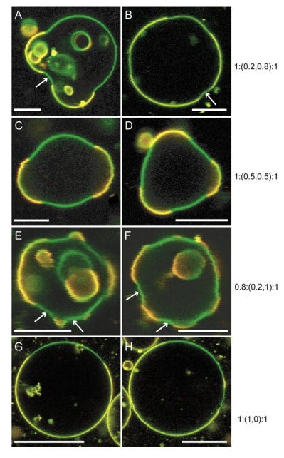
Here are the behaviors and visual appearances of various different design of vesicles as examples for comparison again. The material is definitely of these classes or types. If you have been following then you know that I have been showing these exact structures and behaviors in far better quality than most microscopy in the available literature. Firstly one of my images of a loaded multi-lamellar vesicle in blood, my blood. Its also known as an air bubble if you are a complete idiot. These no longer can be hidden.

Examples of vesicles and some papers for perusal, below. I have folders of these papers and they differ in their constructive properties greatly. Built for the task in hand these kinds of things are what a smart person would refer to as playing with god, or trying to be god himself. Synthetic cell biology is definitely not one of mans greatest achievements, the bad guys like to program whatever they want in these kinds of constructs and it isn’t all good. Specially when you are sticking it into someones body and you have to trust what it says on the label, or what the Dr thinks he/she knows is in those products. It is just pure insanity. OF course, he/she wouldnt know, nor would he be able too what was in said needle or on said swab. A trust for which I would never give to corrupt industries and governments, or just anyone ever. But so many did and still while having no idea what this kind of technology could do to them if in the wrong hands, and that is whats happened. People think its all advance nano medicine, they have no idea what you could put in these things and what they can change inside and outside of your body.
The technology exists to integrate man into a machine, of custom altered design, and to allow technology to be inserted that you never knew existed until the plandemic began surfacing papers and nano concepts from days gone by long ago.
Click image caption to visit paper: just a few examples which exhibit familiar features.


In blood I have been observing all traceable similarities or same phenomenon. Here is an image of a vesicle in my blood which I was mapping the assembling contents from. I wont infer anything from this image since I do not see it provided useful data on its own, but it is showing us that these are again multi-lamellar vesicles with material self assembling inside. Just as we see on COVI-19 RAT swabs, anesthetics and in the vaccines.

Lets look briefly at the just a few of the other nano materials for which many are not able to see clearly enough via dark-field to detect the unique features.
The image below shows a range of different CNT’s surrounding the crystal. The image is taken from Omicron BA 4.5 again. There are multiple colours of CNT, in various geometric shapes, and with differing colours.

The Image below is an over enlargement showing 2 different colours in the one frame. Some CNT’s can look very different and even have segmented colour arrangements, which is a significant observation.

From the same samples we can see some self-assembled materials aligning with the sodium crystals. I am not sure this is intentional because of the sample not being in a biological target and because of how salts will reacts with these materials during saturated assembly environments. It doesn’t seem to happen very often in blood at all, and instead of seeing these kinds of crystals it would appear most of the material is bound for our cell structures.

An interesting fiber shot where the phases can be seen transitioning from snotty like liquid material in to a solid polymerized structure instead.

The image below shows a cut out metallic structure which had formed with others. The first image is the original image enlarged. The image below is using Enhanced image up-scaling software.
The two match very closely without any de-noise, or sharpness added. Just up-scaling by 60x the original image resolution before enlarging it again. One must admit, it is rather impressive and quite accurate looking between the two. A keen eye can see the image is just clearer and sharper, even when enlarged it is clearer than the original. I am unsure of what these metals are right now and there are various types which form. This may be iron related? time well tell, when i can find and afford the right spectrometry kit.

Just as it is in the blood and other products the Omicron Ba_4.5 provides us with well matured multi-layer vesicles constructing other material inside, including what appears to be more Hybrid-LNP’s.

Thanks for reading. thanks for supporting. Its a bit vague when i wanted to show of some of whats going on here, but I am documenting stuff and probably shouldn’t be spending too long Substacking whole long topics. Best it gets organized and place in a tree system on my website. In the mean team enjoy the food for thought and please do consider buying lots of KO-FI below so we can all see detailed spectrometry results from purchased equipment. Microscopy is great, but now it is time to target each structure to learn specific compositions and purposes. Wishing everyone well, apologies for rushed type and neuro-mistakes.
Ill leave you with a recent blood image!


Google Gmail blocks content from your mailbox for controversial material or websites. I encourage all freedom type folks and awake people to avoid gmail for this kind of email subscription. they are terrible for it right now and i am getting many emails complaining of not getting posts or being able to send me email. Ill bet you have gmail ! do mention if not. Thanks!
Well you have been incredibly busy...what an amazing labyrinth of detailed detections you have
compiled herein, seriously Karl !! This represents an invaluable resource for many of us trying
to understand just what exactly we are looking at in all of your breathtakingly beautiful imagery
you've been spoiling us with. I am sure i am not alone in really appreciating the degree of painstaking study and sorting-of its taken for you to have organised this impressive legend.
I really appreciate, and wholeheartedly agree with your conclusive remarks, as to the evidence of contents uncovered speaks clearly for itself = as identifiable and identified ! ......and not ppl' s wild imaginings we keep hearing is the case. It's such an insult to intelligence, and at how incredibly lazy to not give this poisonous blight on us - its due examination.
Incredibly thorough workout, marathon man Karl !! Appreciate "your" due diligence enormously, KK
Thanks KoppyKat.