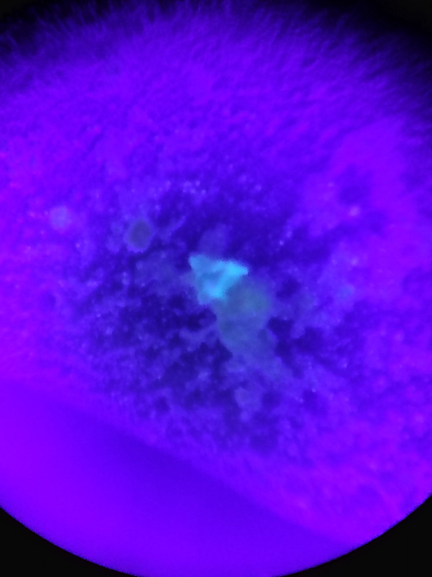
Stool sample shows UV reactive material and Q-dots.
A look at the strange structures using Ultraviolet light and dark field microscopy.
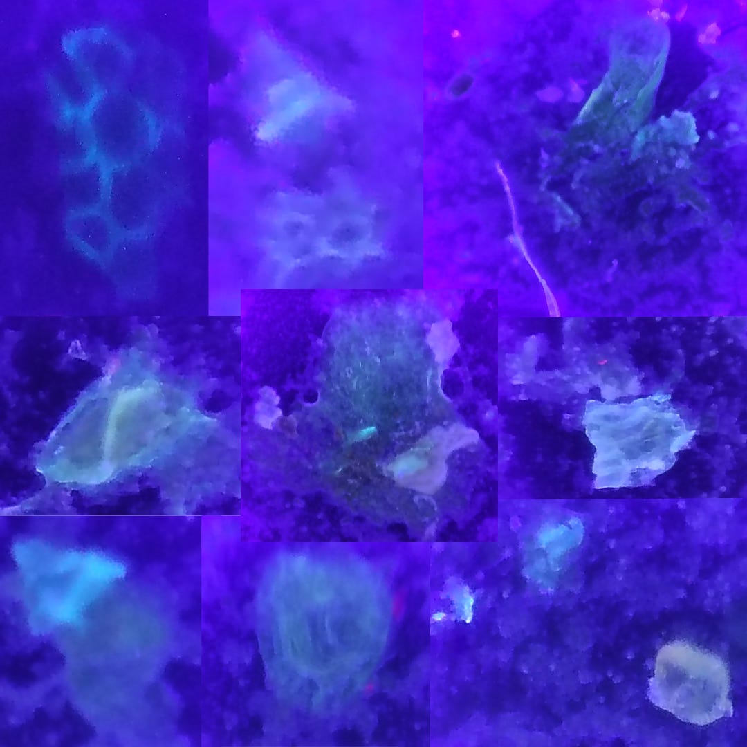
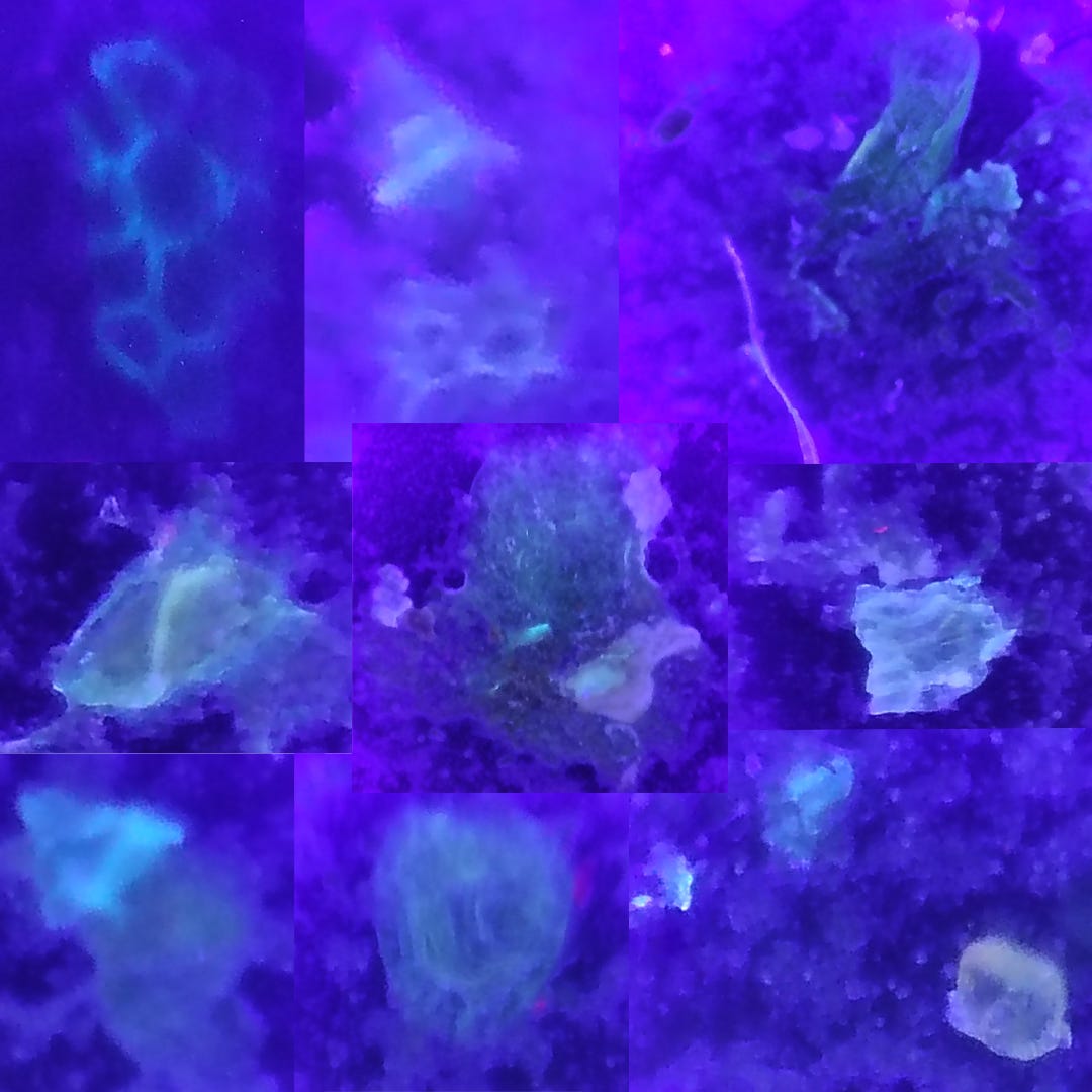
Above: UV light used for stool sample. 9 images collaged together in photoshop.
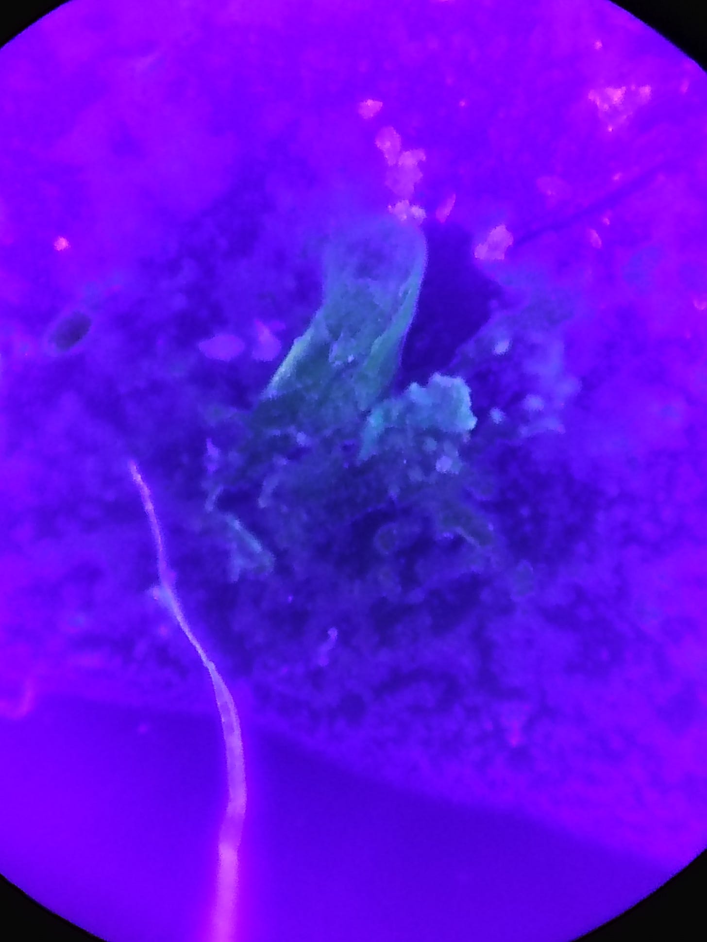
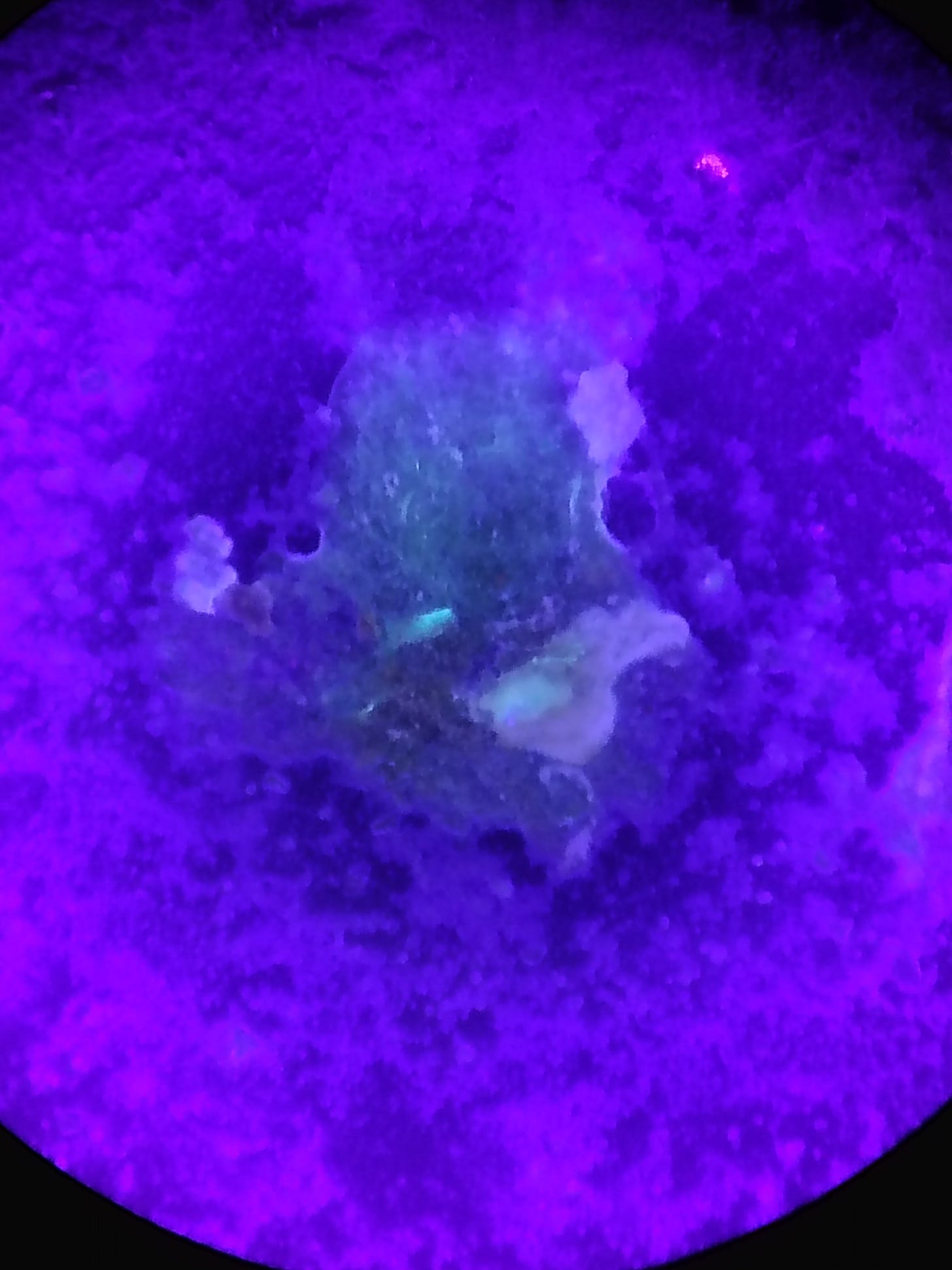
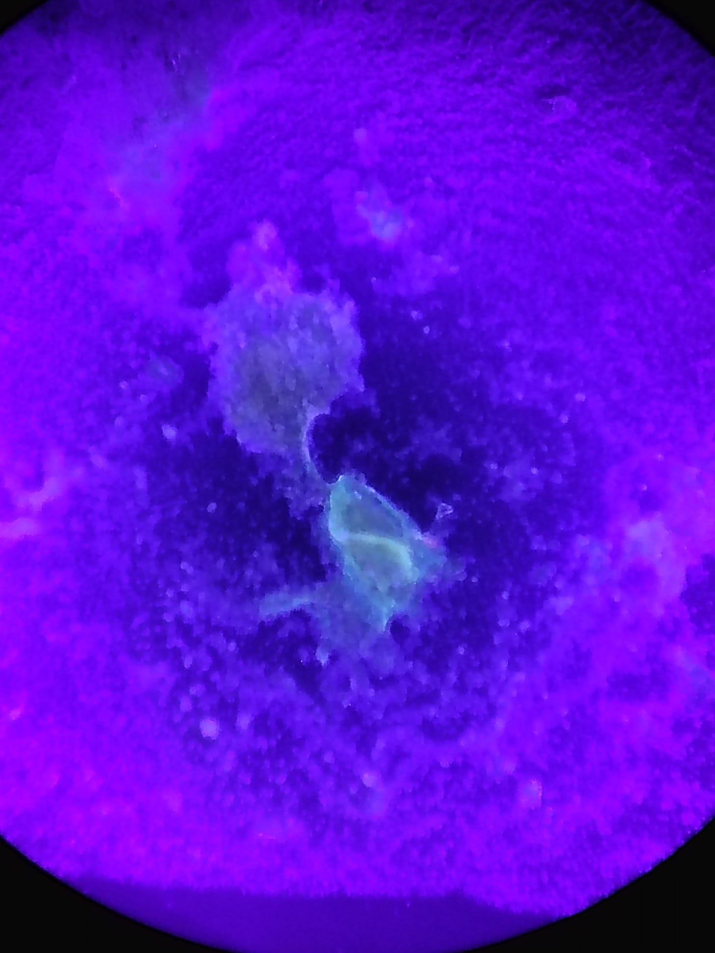
The image quality could be better but unfortunately the microscope camera has a part UV filter which only shows some structures. A phone had to be used to capture these. Amazingly some of the wavelengths excited by the UV light simply left the structures completely invisible to the microscope camera.
Some of the fibers also illuminated very brightly, along with other gel structures. I could see plant cell structures which appeared normal and just to be sure started testing plant foods using UV to make sure these materials were not those. Certain Fungus is often UV reactive and shows this same yellow green reaction. I suspect that whatever makes these structures is still partly a fungal organism of the Dictyostelium kind possibly.
While spending much time previously observing morgellons samples I could also see no end of These glowing patterns and strange fruiting body like formations. I once took saliva and grew a whole ferraro rochae tray full of dictyostelium slime mold from a saliva sample. Most of the small fruiting bodies and masses it seemed to create all had this same appearance in many ways. Of course what we are looking at here may or may not be directly related, only DNA sequencing, and other analysis could help there.
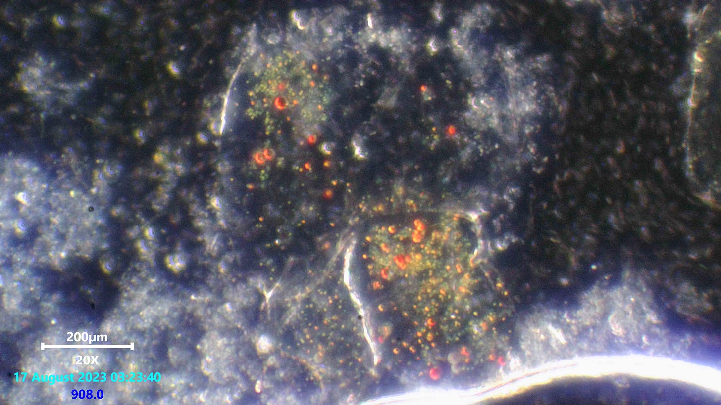
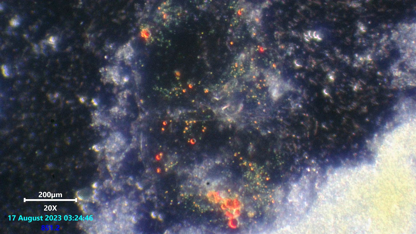
Above: Q-dots with much larger round bodies housed within a gel structure in stool sample, 200x dark field.
There were several structures filled with dots like these in this thickly smeared slide covered with a covers slip. The dots do not seem to react to UV at all so far. The separate structures only seemed to do it on this viewing. The larger red bodies are of interest since they are not normally seen when I find Q-dots in samples.
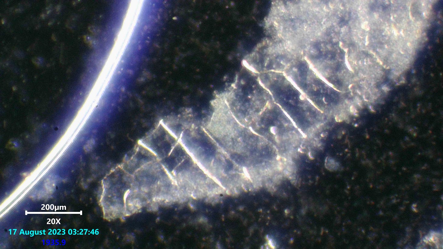
Above: Gel structure in stool sample, 200x dark field.
Again there was no shortage of oddly patterned crystalline gel structures like this one. There seemed to be organisms inside the clearer gel panels trapped inside and buzzing around. They did not seem like any of the other bacteria swimming loose in the sample.
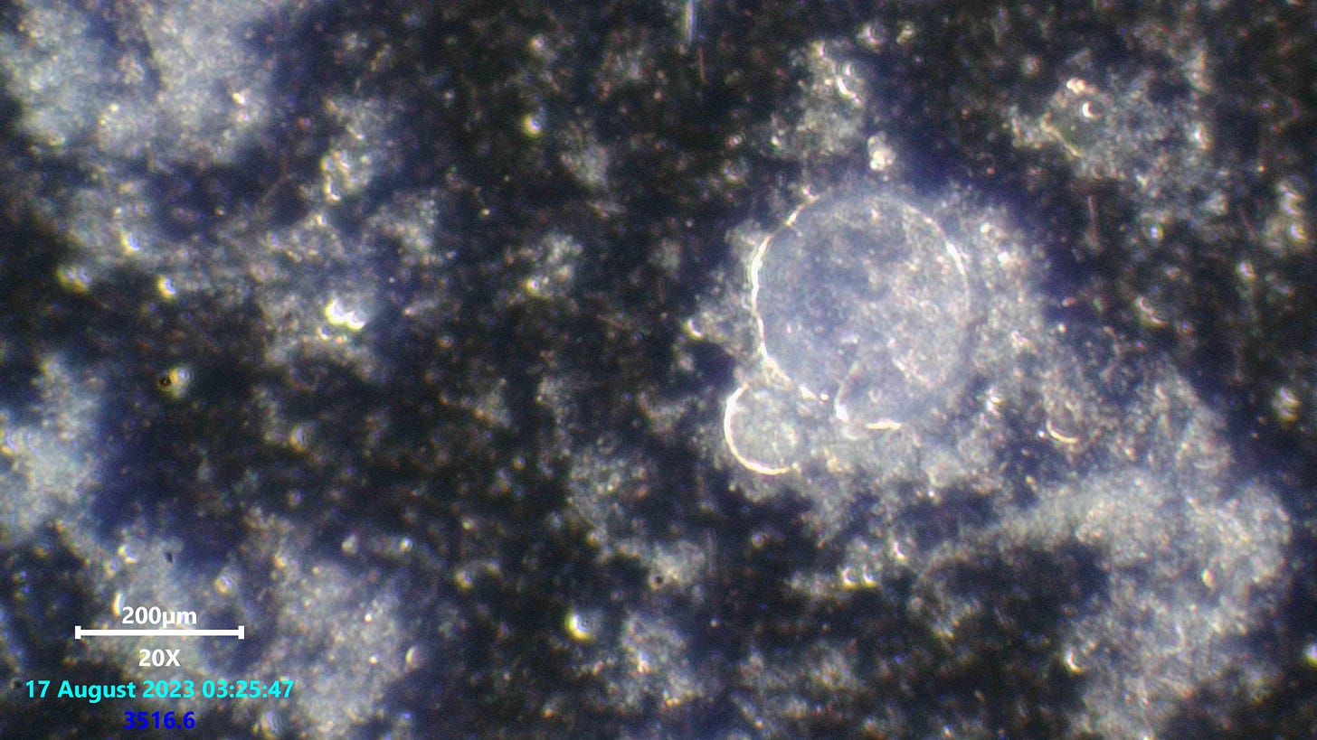
Above: gel structures in stool, 200x dark field.
Again, there were many circular structures among other shapes. All very peculiar to say the least. What was also interesting was the large masses of clear gel spread throughout the sample all over. UV reactive liquids that reassembled the same glow as the solid structures could be seen spreading towards the outer edges of the cover slip. There was a large concentration of this glowing liquid.
Above: another site which had Q-dots.
It seems that all though these dots can often be seen spread through out blood plasma and other liquids, it is often always found bound within gel structures regardless of the concentration in surrounding fluid.
Here are some larger images from the collage for better viewing.
.
Help the citizen research by helping to provide equipment and supplies so we can go deeper.
I am using KO-FI right now and appreciate you clicking the link to go there and contribute.
Thank you every one !



Good old fashioned original work. Hard to find these days, cheers.
More please.
Incredible work, Karl. Is it possible that this gel you are seeing is a form of biofilm, which is generated to protect these things?