


Worlds most powerful dark-field microscope ? It would seem so.
I could find no evidence anywhere of a microscope having this much Light power, neither could chatGPT or google. It may well just be the first of its kind.

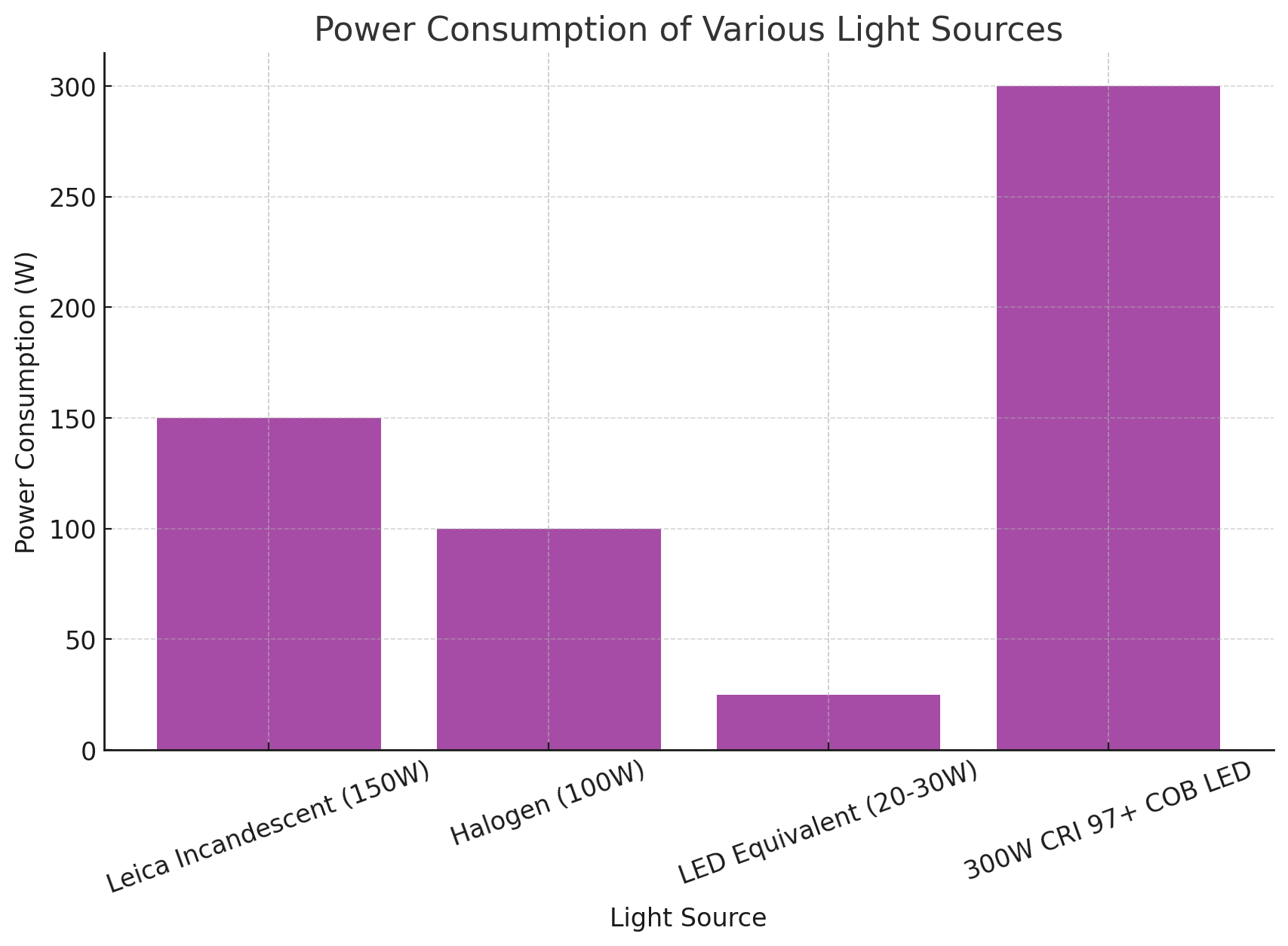
Exciting !! When I ordered my last 100 Watt broad spectrum LED I also ordered a 300 Watt version. What I forgot to order was the power supply needed to run the 300 Watt LED and so I stuck in with the 100Watt COB LED for a bit and it performed exceedingly well, but not this good. The optical quality is shocking. I am going to share the first day with you.

The light system is absolutely working as expected and currently it isn’t even a corrected beam exiting the COB LED. The correct lenses to converge the multiple LED’s on the COB and refocus the light beam at the condenser have not yet arrived. This may offer a small amount of contrast improvement, lessening some noise and increasing the sharpness of outlines in more detail.
The LED has to be mounted on a massive IGBT heatsink and with multiple 240v industrial fans. A 60v 7Amp switch-mode supply is powering the light source through a variable controller that features current and voltage monitoring. The base of the microscope body still gets painfully hot to touch, but funny enough the original Leica light source Glass remains fairly cool. The heat reaching the slide is far lower than expected. However, I shall still add a small turbo fan to cool the COB LED from the top side where almost all of the heat not taken away by the cabinet and heatsink fans ends up accumulating the most. From there it transfers heat into the scopes steel body.
One look at the LED directly is painful. High-power LED’s can be very dangerous indeed and should definitely be considered as a high power laser when being handling. LED’s tend to produce more collimated light and include more Blue spectra than most filament bulbs. They are definitely closer to laser where safety is concerned. I didn’t even look at it, I slowly turned my head towards it from a distance at half supply current and instantly felt discomfort. I had to shut the casing panel to even face the same direction….Fun! It needs a few more tweaks, but the results are already super impressive by all documented microscopy standards, detail is seen within structures at yet again new levels of image quality. The colour is suburb depending on the objective used and the resolution has definitely reached a point where the only improvement we could see from here would be to upgrade to an extortionately priced imaging camera. The one I have is already very impressive, the rest of the setup has reached its maximum potential. I cant afford another camera now since the new FT-IR spectrometer needs service and a few pricey parts before she is running. The spectrometer is my priority at the moment as it will help us confirm some of these materials using multiple methods. All help is still greatly appreciated with KO-FI donations and I shall be sharing ALL data that comes from my work in appropriately documented form. Back to blood business and what the new setup is achieving.

Above video: A little further out we can see slightly less developed RBC’s which are forming materials inside slowly. A sea of Blue particle bodies are excited by Brownian motion, optically they appear around .5um to 1um. Due to plasmonic effects the light we see is larger than the actual structure itself, the particles if seen to be .5um are likely far smaller than that and definitely in the nano particle size range. As time goes by we can see the particles are assembling constantly and it by day 2 it is evident that they are synthesizing more of themselves. Either from non-visible nano-materials in the plasma, or from the large Bi-Layer vesicles (Yellow whitish structure in the middle). These Bi-layer vesicles would effectively act as complex bioreactors or chemical factories. As the vesicle breaks down from environmental destabilization it leaves visible clusters or strands of complex material which is believed to be complex building blocks of the almost intelligent membrane.
Above image: Taken at 20x using a LEICA CORR (colour corrected) infinity objective, Still my unvaccinated blood. Iridescence shows beautifully in some RBC’s, that seems to happen as a stage of transitioning or again environmental destabilization. Strong iridescence is not generally noted with natural RBC’s as much as it is pronounced in synthetically altered or coated RBC’s according to literature so far as I could find. At 20x we cant really see much and the blood might even look almost fine to some, of course the giant bi-layer vesicles are reasonably obvious still and cant be confused as bubbles or gas, even at 20x magnification.
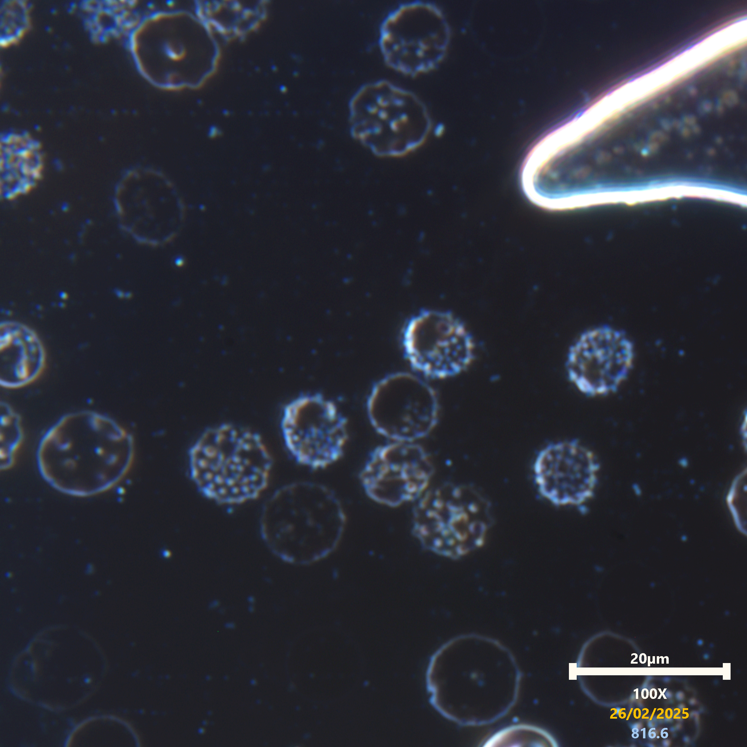
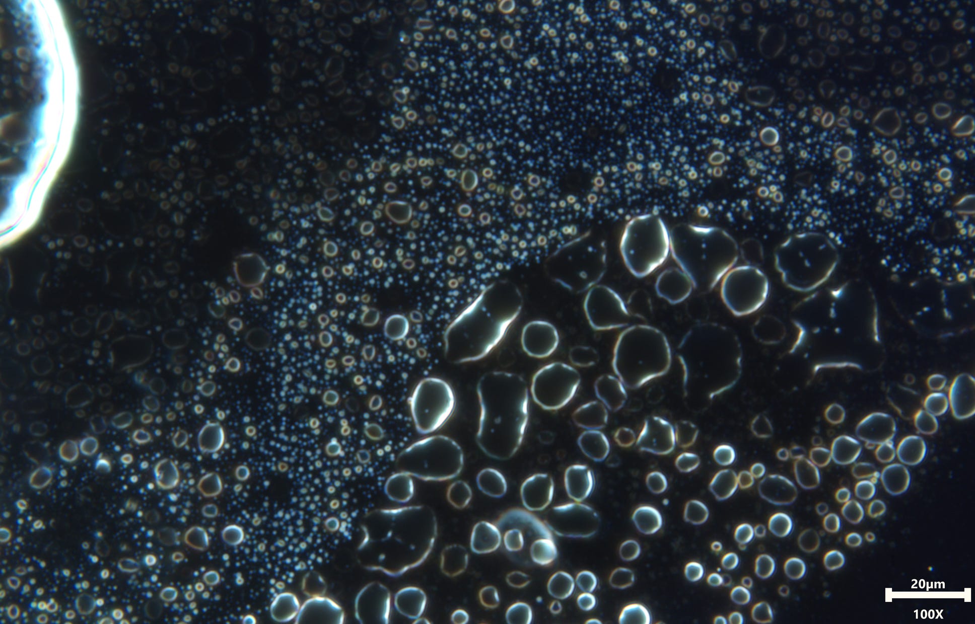
Above image: I shall mention now that all the images in today’s post are my own unvaccinated blood, that’s easier! This image at 20x is at a different location of the sample where the Vesicles were loaded with material which forms over time. The material appears to fit the behavior and morphology of Hybridized-lipid-nano-particles with polymer-like features. Some of the vesicles formed with outside pockets inside and we can still see the RBC’s in the center. This image shows that even using 20x magnification it is possible to clearly view the internal processes beyond that seen in previous light source configurations that I had made and tested before.

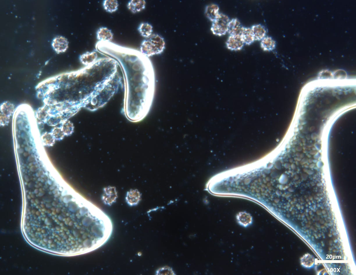
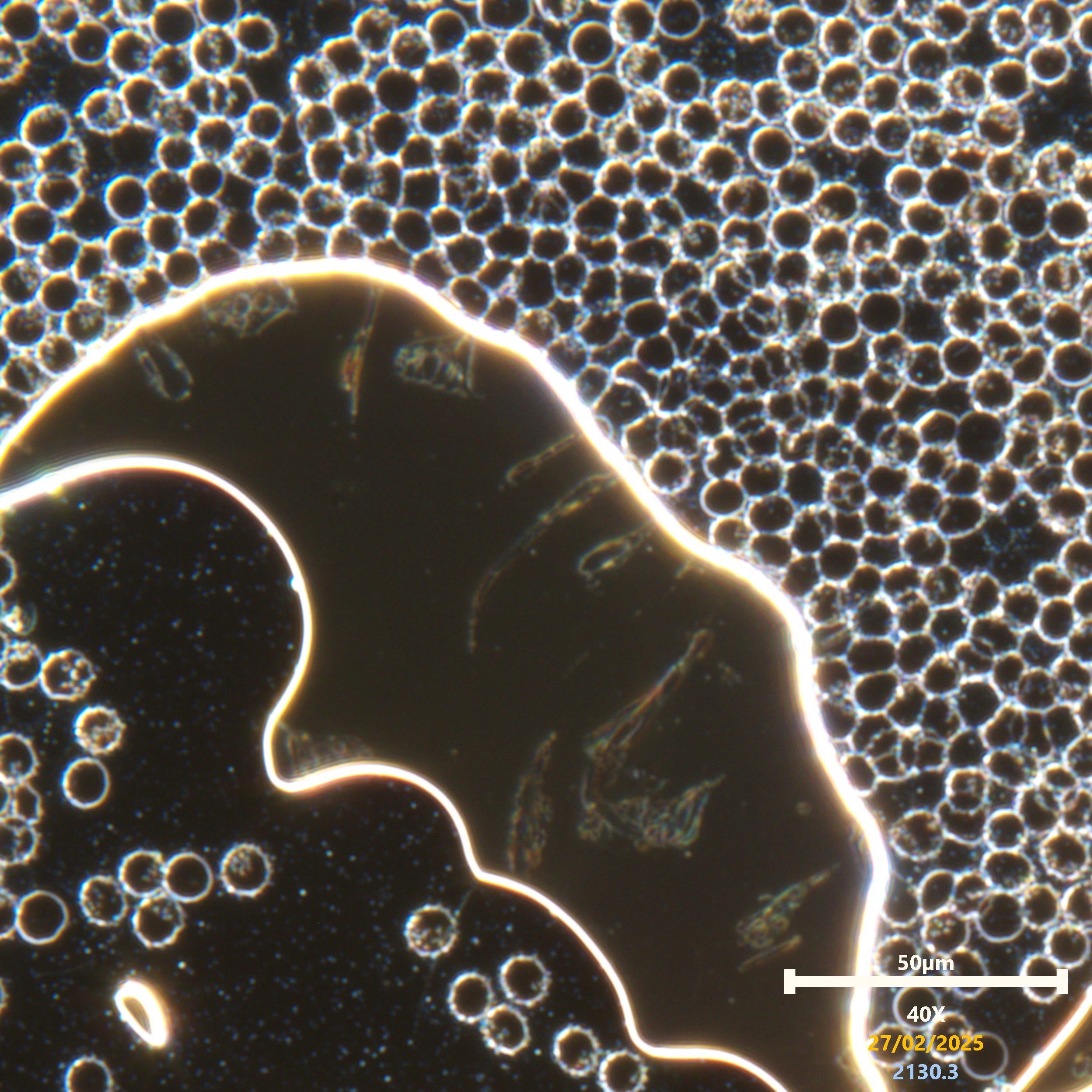
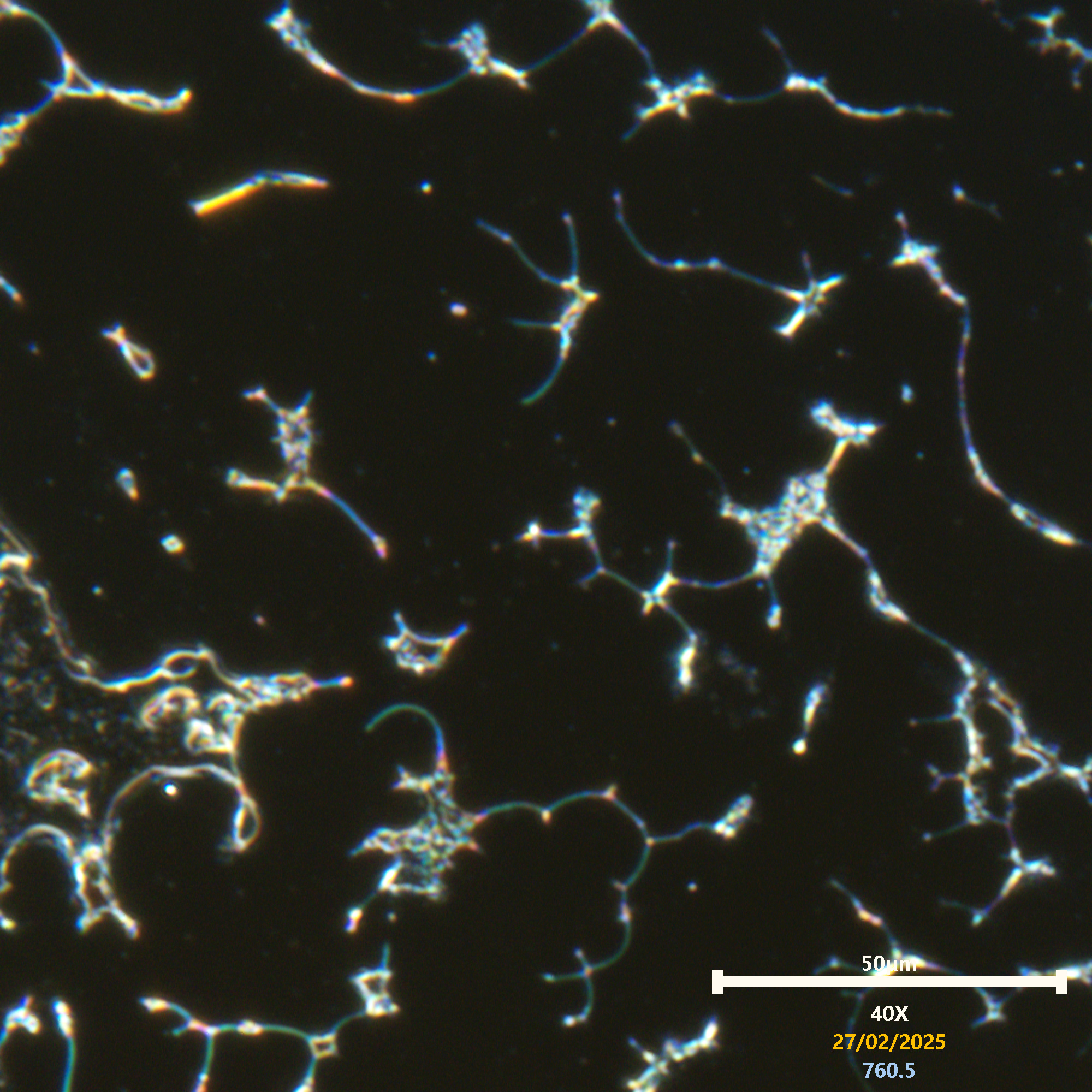
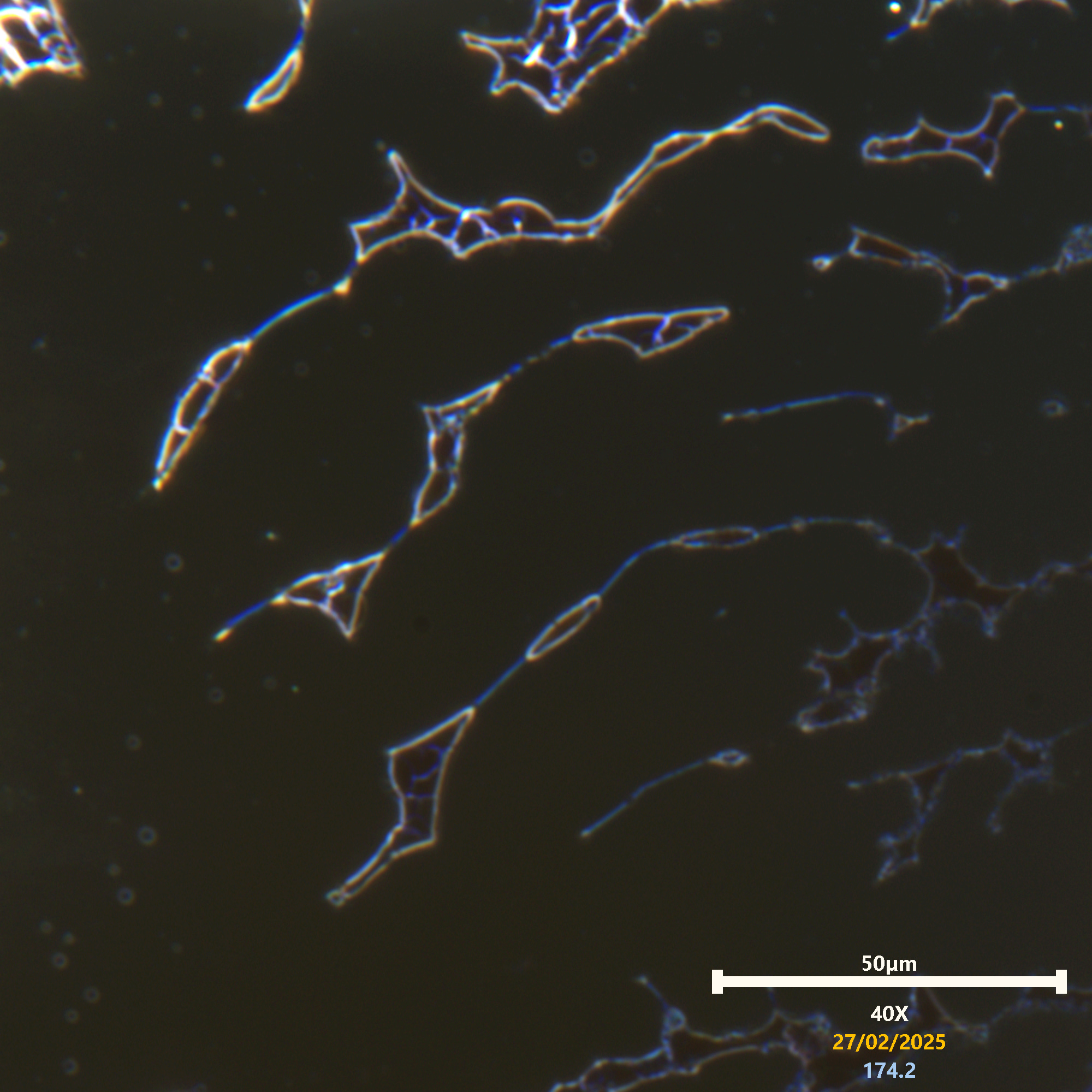
Above: 40X magnification has the best balance of aperture sizing for the light conditions. This objective gives me greater resolution and colour than the rather expensive 100x could ever achieve, but it also has its limits when enlarging a frame. Again, we observe the strange polymer-like presentation of the RBC’s, clearly not what should be expected without some kind of significant membrane alteration occurring. The give away for me has definitely been the materials I have been observing assembling inside the RBC’s for over a year now. Put the two together and you have something which can only be explained by the DOD’s Erythromer artificially altered RBC’s. Proving this will need the spectrometer running, and some other analysis techniques to be performed. Did I mention that KO-FI or any donations towards the Spectrometers service would be beneficial for us all!
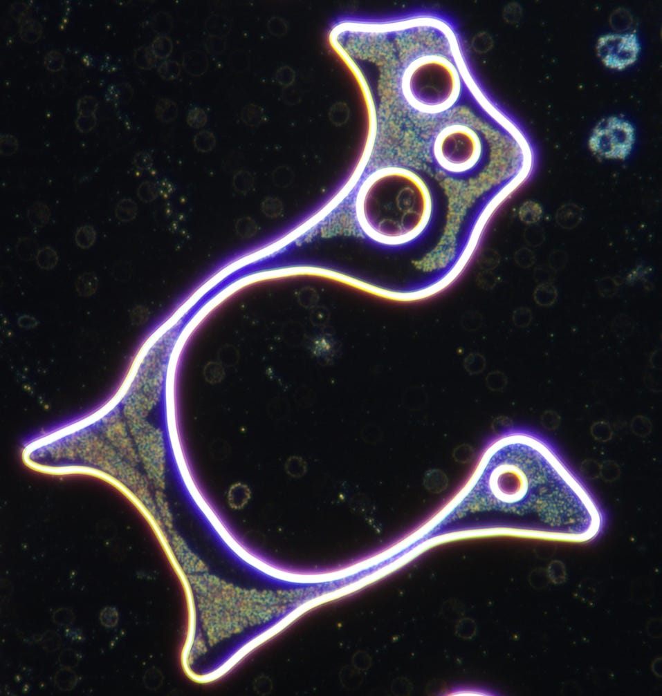
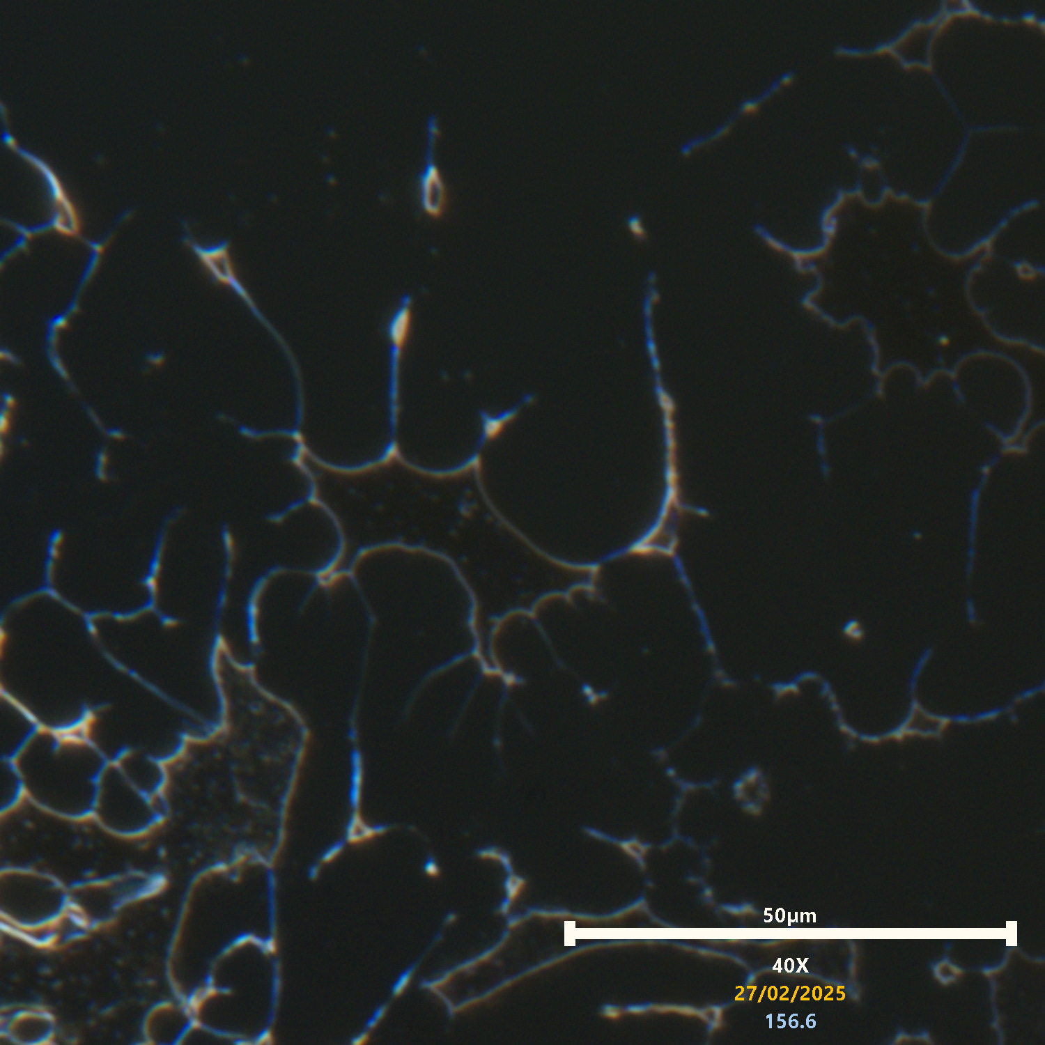
Above: Another absolutely stunning image of lipid-like formations developing inside of the Lamellar vesicle. The vesicle exhibits hugely bright light wave patterns as a result of the material types in the membrane. A lonely polymer has solidified, has various exotic particles, crystalline structures, and likely other biological materials embedded within. It is worth noting that the bright materials within are mainly seen to develop within these solid structures and not so much seen with the other foreign materials identified in the plasma. Those particles and structures seem to have very different appearance and morphological patterns. Some RBC’s are clearly showing the gel stage before reaching the stage where material forms uniquely after.
Above: A striking glimpse into a developing vesicle which has caught the lipid-like form of the expanded hybrid-nanoparticles. On occasions you can see that some smaller blue particles will bridge between larger lipids, a known behavior of lipid-polymer based constructs which can behave just like this.
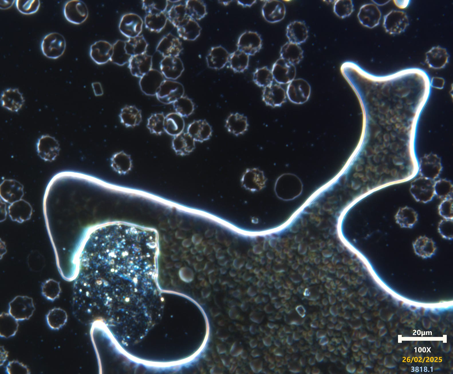
Above: Excellent 100x capture of the particle maturing in more variety of stages. We see the small blue particle expanding into lipid-polymer vesicles towards the end. Some of the larger vesicles on the right seem to be made up of multiple smaller and much more colourful lipids that reveal differing colours as if some are unique to each other. This may not be the case since a structure of the same material may produce different colours depending on how light waves behave inside the structure based on what its made of. From what I have been observing, it is likely that there is unique design between various lipids.

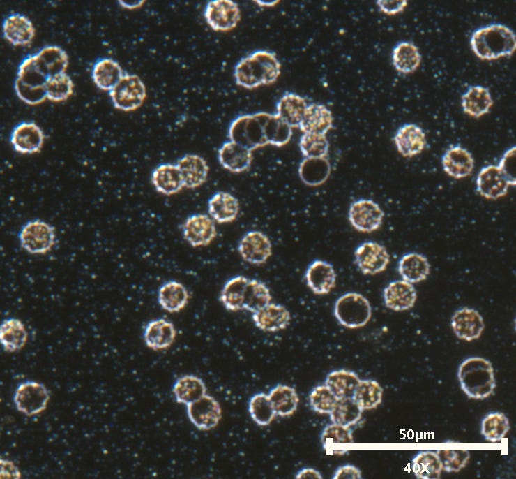
Above: Variations in RBC’S as seen in earlier images here is a vesicle and liposome-like or exosome-like forms just above which used to be considered pleomorphic bacterial forms until more recent studies showed that there were no bacterial indicators present or evidence to support the pleomorphic theory.
Pleomorphic Structures in Human Blood Are Red Blood Cell-Derived Microparticles, Not Bacteria
All other previous studies were without tangible evidence, conjecture based, or poorly carried out with contamination not factored or considered. Controls were not performed in the more convincing studies either. The two studies above DID use correct methods and approach to making their claims, they discovered that positive bacterial results came from contamination and correlated it to what was seen in blood. This is relevant to this work since liposome forms are present and they seem to exhibit synthetic features. With all the previous questioning of chronic disease one has to ask if similar structures seen there were also synthetic or if they were actually natural lipids.
Above: Since we have discussed most of the phenomenon in this image already I shall reference only the colorful smears inside of the vesicle. These used to appear very faint and featureless, they now exhibit bright colouring and are more visible than ever before. What these are I have not yet figured out. This is also something you would of seen more faintly in Lyme or chronic disease from microscopy in papers. They used to catch my eye and I would wonder why there was no attention payed to some of these disease related features. These obviously have the hallmarks of unusual synthetic material, but maybe not.
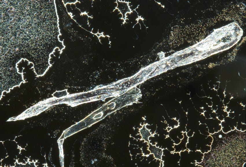
Above: a fiber in a stitched image at 40x. The fiber is highly responsive to moisture and on occasions the phases can be seen changing with the hydration varying in the structure (bubbles moving back and forth). This is also a notable behavior of more complex synthetic fiber materials often associated with wrongly claimed “bio-compatible” hydrogels. The fiber is laden with varying materials, particles, coloured streaks, and other bizarre structures. It is unusual for a simple or natural fiber to have all these features. More coming on some of these separate topics soon and with references to explain or support the points for which I raise.
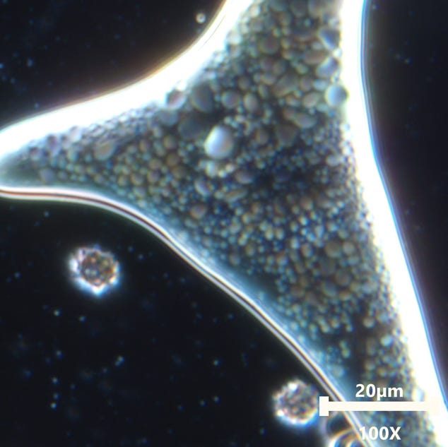
Above: An enlarged image taken with the 100x objective. Inside the blood cell there are sophisticated looking structures. It has been beneficial to hair so much light power because it enables me to turn the gain down and achieve much higher video frame rates in dark samples. Remember that the 100x isn’t as colorful as when using the 40x objective, these structures are much more colourful than they appear here..
It is getting late and I think that since we have covered mostly what I am showing in these particular images that I will post some more videos and images without commentary just so we can see a bit more on showcasing the light source.
These two videos only are actually 40x and not 100x. I must of forgotten to change the on screen sizing parameters, oops.
I shall stop there since I think we have done a reasonable job of exploring the modified light source capabilities. Shame I couldn’t patent it, It would of been nice to have earned some funding off of it for further research here. Anyway, thank you all so very much for reading and watching. Thank you so much for everyone that has supported or helped in any way so far, and thank you for those who may support the efforts moving forward. Please hit the KO-FI page if you want to help out with getting the FT-IR spectrometer going and calibrated to spec. Until next time folks! Nite all.
Thank you Amy!
Karl, you are producing the clearest images of the fungal/graphene payload ever published.
The clear path forward is now obvious to me.
Vaxxed or not, everyone is inhaling the chemtrail fungi.
Their wholly owned MSM tells the gullible masses that Geo-Engineering is a conspiracy theory, believed by paranoid fantasists.
They feed us, the paranoid fantasists/ informed minority, a cunningly devised load of codswallop which is... WE, YOUR ALIEN MASTERS HAVE FILLED YOUR BLOOD WITH RIDICULOUSLY CLEVER NANO SCALE MICRO ROBOTS AND YOU WILL ALL BECOME OUR SLAVES AS TRANSHUMAN CYBORGS. The truth is very different, however.
It is just another robbery. A transfer of property and wealth from us to them and their underlings.
We are now witnessing the early stages of this GREAT RESET... the Hawaiin DEW fires gifting them the most sensational island real estate on the planet then the other day, they did the same to the Hollywood Hills. Mel Gibson, the dissenter gets burned to the ground but Tom Hanks, the team player stays home without a care.
In the meantime, we are all getting sick from the fungal graphene, giving their wholly owned Big Medi/Pharma Juggernaut it's greatest ever boom times.
I have personally witnessed this phenomenon as I nursed my wife through a vicious cancer, then I got a Merkel Cell Carcinoma.
There is no stopping their depopulation agenda and the inevitable transfer of wealth outcome.
We are all passengers.
Under 1 billion humans by 2030 and only the smartest and most resilient of us will survive to see what they will do with Planet Earth.
I am now strangely content and relaxed about it all.
BRING IT ON.... PROTEST IS FUTILE.