Alert! more on the nanotechnology and biological delivery system's under study.
A new method of viewing the swabs has been discovered by accident. Lots of images to see!

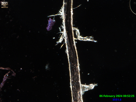
The colorful dots which are varying Nanoparticles (NP’s) can clearly be seen. These and other material are in fact formed mostly by the lipid structures seen littering the slide, It seems there are already formed NP’s being found in other sources such as utility water too.
All this can now be seen with more certainty thanks to a new technique used to observe the material unfolding for longer and with increased optical clarity. The NP’s do not create the lipids as somebody else suggested, they may carry material on their surface spreading more, but these seem like they are initially created by a bottoms up approach inside lipid or polymer encapsulated cells. This may not be the way it happens much later on when it wants to produce more in different environments. It seems there is an unpacking process of the original product and then a different set of productive or self synthesizing stages in blood. It is too early to draw conclusions on how that bit works precisely yet but our own cells seem altered when viewing blood.
For this sample I used 3 Dye’s following literature on Hydrogel identification, Fast green, Alcian blue, and Congo red. In this case the intention was to indicate the presence of particular Hydrogel’s and materials suspended in the contained matrix. I will post info separately.
While I was playing around with combinations of swab and buffer I realized everything became far more extreme and chaotic. Everything was reacting insanely fast and things seemed to be sped up. I figured adding the cover slip will slow it down after letting the first series of reactions take place. We already know that both the swab and the injectable’s require oxygen which we believe it would usually get from you and at much lower exposure than out in the open. Lets not also forget that it is highly light responsive material and the amount of light has affects on how fast the material reacts as a result.


Above we can see that there is a Hydrogel like product that forms on the outer of the main solution droplet and seems to play a part in forming polymer based encapsulations. Some of the polymer structures do not stain or sometimes take up a different dye colour, this implies they are different polymers. Dots were rushing around inside some of these capsules and reactions were occurring within, some with particles or other cells moving around. The middle image shows various materials. In deep red there seems to be the part formed gel responsible for the polymer cell structures which later still maintain some of this dye colour. There are also solid polymer structures of a different kind containing bright NP’S and they did not stain at all, these may be PEG based? Still learning the details of this broad topic.


New cell structure morphology could be seen from many of the different cell type materials compared to what is seen via more instant viewing methods. The sample is still under observation, but the morphology has slowed down likely due to having absorbed resources and lacking environmental factors.

What
would label a 10.5 bar beast, He had blood pics showing above crystalline morphology.


Brief summery:
While we can see that this is all very complicated and certainly what you would expect from exotic science engineering, it is also indeed very important that we know exactly what these materials are. The dye procedures are very complicated to interpret without jumping to narrow minded conclusions, so we will take more time, research, and have more discussion between ourselves before doing so.
Some forms of spectrometry analysis would likely struggle to pickup on the wavelengths of material embedded in unpacked morphological structures accurately or without misleading without proper separation. I think that in order to get things right a combination of dye’s, Visual observation, and varying forms of separated method spectrometry will be key to triangulating the ID of components in the mix. until this is done no one knows what we are dealing with. It seems that the dots are complex NP’s (not robots), there may be robots present at later stages that look different to the coloured dots or are much bigger and some are of non-rounded shape. The lipids and other structures can move in culture, but they are not robots just because they moved.
We can learn a lot from dye’s and see how SOME of the base materials end up forming. There are things that we will not see happening using microscopy also and those things are likely on the Nano scale. I shall continue playing with these dye’s and a few new dyes before I draw any suggestions to which Hydrogel’s have been used.
My thoughts at this point are that a 4-Arm cross linked PEG based Hydrogel product is in use. I have managed to see 2-3 possible gel’s present via specific dye technique’s. Common PEG arms can be Chitosan, Alginate, Calcium based, Silk, and more. Materials loaded into the cargo or matrix can be biological and non biological such as nano-materials, some of these materials can show up using various other dye’s.
I appreciate all the support so far and urge those who can help me continue doing the work. Soon the tools required will become much more expensive.


Hello Karl, I have what might be a terrific idea to sponsor your important work.. Can you offer a service where you test materials sent in to you from your readers and the public? For example, IVERMECTIN.., lady's liquid face makeup, store bought products - especially fluids. ? You charge for the service and we figure out what to avoid. What do you think?
Two things that may clear the blood. Chlorine Dioxide and DMSO. Both have been around forever. Both have been heavily censored and vilified. You will need to use search engines like Brave, Rumble or Brighten.