Different perspectives of microscopy exploit possible details of interest.
Material in blood may possibly exhibit same light wave patterns in areas containing some of the questionable materials, or areas of blood that have been damaged after being coated in such materials.
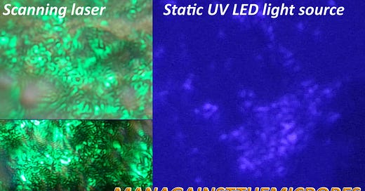
Images to left are of previous scanning laser observations. Images to the right are images taken with UV light source approx 410nm wavelength.
The same light wave details are observed on gel like formations also recently described by Shimon Yanowitz as possible being proteins. He may be right there, but it wont be long till more is known. For now think of gels and proteins as interchangeable with our current lack of understanding. The patterns seen here are light waves bouncing off of textured walls, divides, creases, dips, raises, etc. The resulting pattern can be quite telling, more so that we can more distinctly link the locations of such material in blood quite easily.
Remember that in previous microscopy videos we showed you this stuff moving at some stages, and that this could not be seen between RBC’s using bright or dark field microscopy. Lets look at some of the other suspect artifacts in the blood which exhibited this same pattern. Bare in mind many other structures of which had different features such as colour, shape and size did not exhibit this same light pattern even if the structures looked similar to our suspect ones.
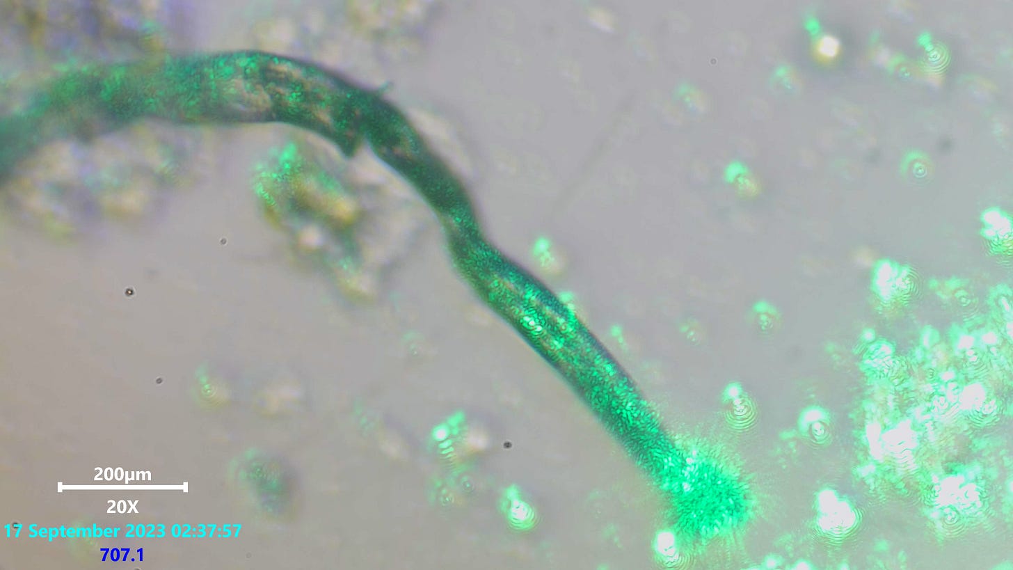
The Fibers exhibited this light wave pattern on some of them, but not all. It was consistent at varying focal lengths but did not appear on many other landmarks or features within its close proximity
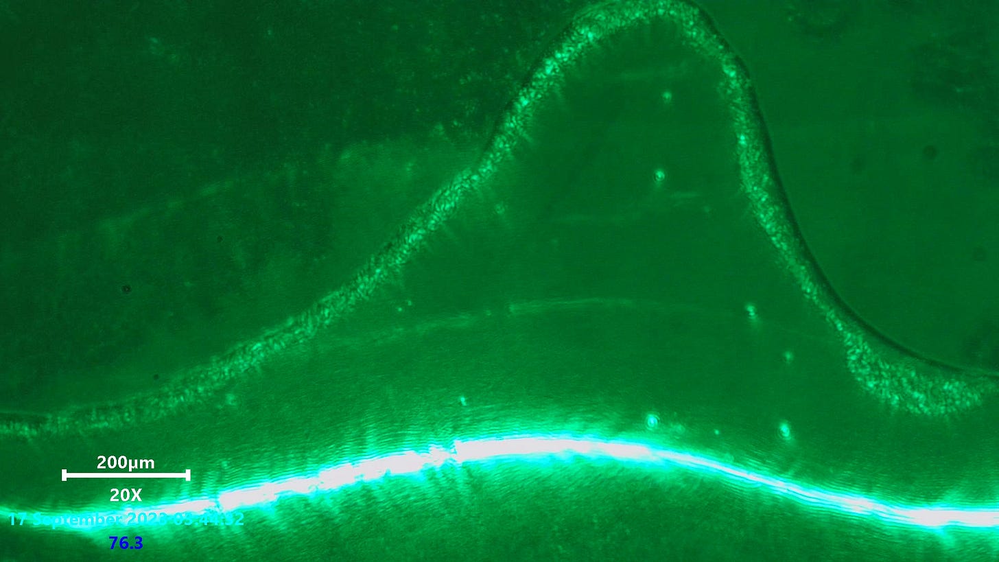
It was seen at the borders where smudged blood met a void or what was thought to be a bubble. This material lined that border in varying amounts.
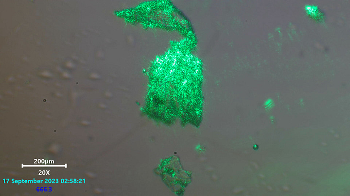
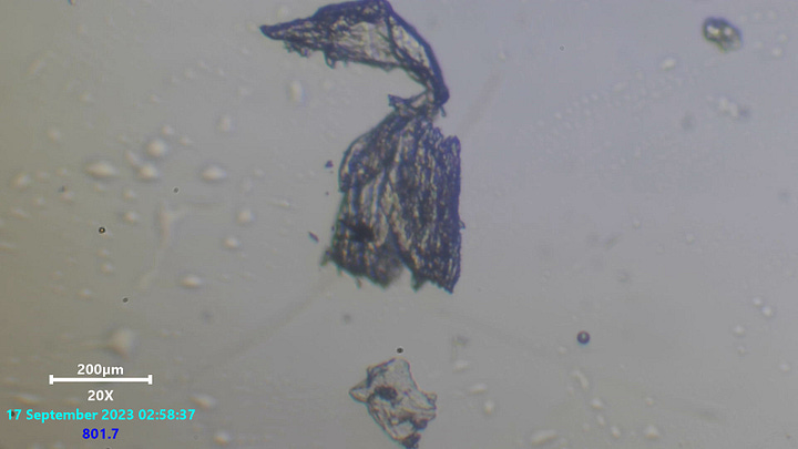
Other structures such as these folded ribbons contained the same textured light effect but was noted to be more as if it was on the outer layer. This might have been mistaken interpretation though. Notice the slide contains other much smaller material which is not excited this way either.
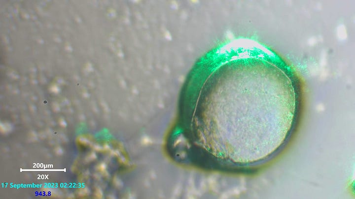
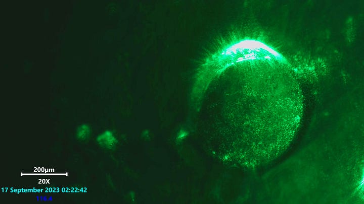
Some of the crumpling spheres exhibited the exact same and distinct light patterns also. These structures are obviously materials of interest where research is concerned now.
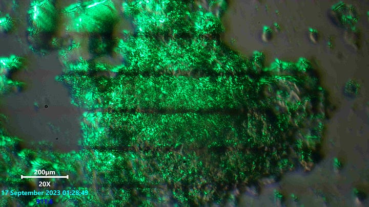
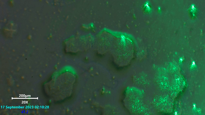
More structures or curious sites where the pattern was exhibited.
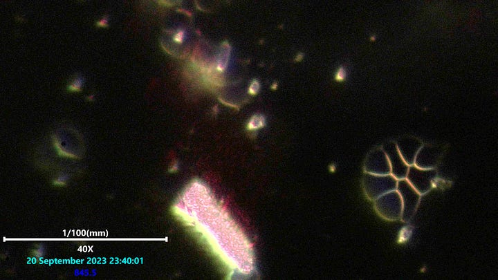
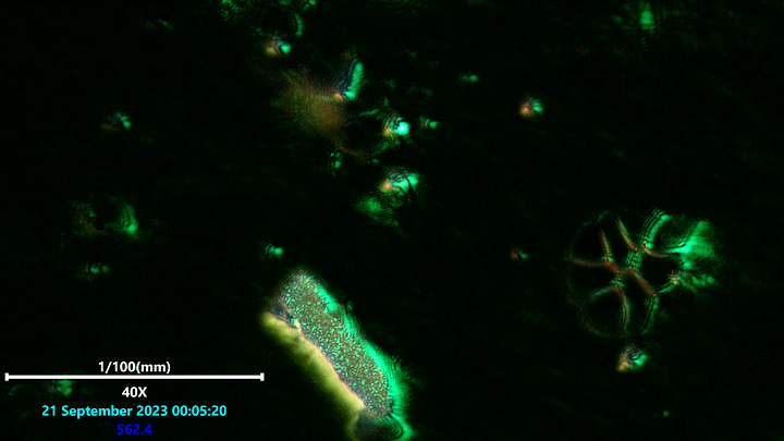
EXTREMELY clear patterns were seen in the mass to the right. Laser on the left was RED and to the right was GREEN. Both in scanning laser setups.
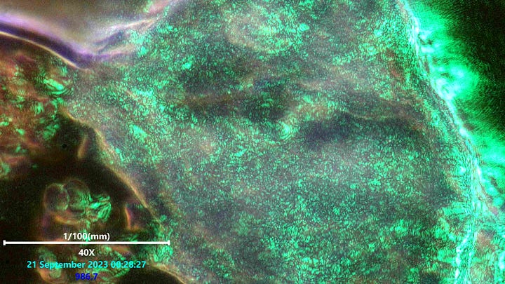
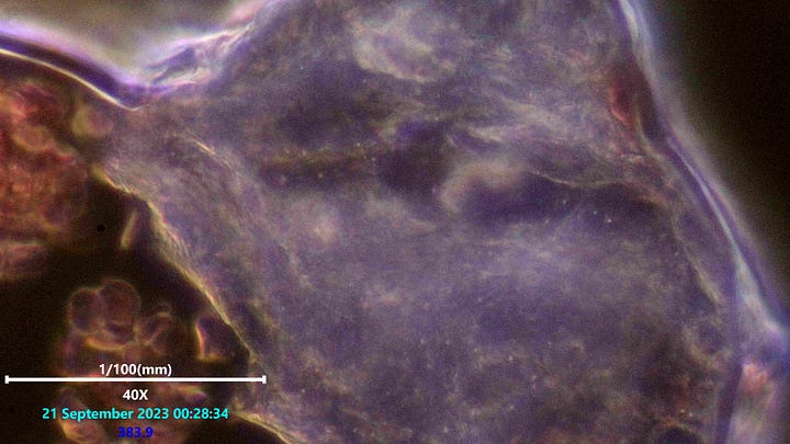
A large mass at 400x next to smudged blood. You can see similar or same patterns here too. It was noticed that in areas of blood where the blood was still in tact that no light patterns matching these were seen in the RBC’s at all. Blood which had deformed and smudged together began showing these light patterns throughout the whole mass of every RBC at that point. Before the RBC’s were damaged and showed no patterns inside it was observed that these light patterns could be seen outside of the cells as if something outside wanted to be inside the RBC’s. All the locations where this pattern was found inside the damaged blood showed no patterns lingering outside or around the RBC’s anymore.
I found these ventures rather interesting so far. Something could be misunderstood here on my part, but there it seems that the above points of interest may be indicating that their is something going on here. Much more light work will be played with before drawing any solid conclusions. But here is where i have gotten too on so far using these techniques. Time will tell as usual.
Big thanks to those who contribute to any of the citizen researchers out there. Help out with KO-FI below.



Aloha to everyone and mahalo for your great work and devotion to science and the truth. ALL information derived from these different lighting techniques is valuable. The ability to adjust the frequency of the laser light might be something to investigate. This being said, my thoughts are more focused on the surface charges used in the self assembly process. While it is difficult to imagine how any particular frequency of light would illuminate surface charges, Kirlian optical methods would do just that. Furthermore, the frequency of the Kirlian high voltage source could also be varied and optically observed. Information theory leads me to believe that the Corona Discharge observed in Kirlian optical methods would make these self assembling nanoparticles and processes more transparent and more understood than they are now. One more thing; We do know that EDTA IV is effective in cleaning up the blood and restoring normalcy. A case could be made for using or oral doses of Potassium Iodide, which is a non-toxic Ionic Binary Salt that can be used to chelate heavy metals and toxins from the blood. this should be done after 6 hours fasting and no food for 2 hours after ingestion, because the actions of the bipolar Ions I- and P+ will quckly be spent combining with minerals and nutrients in the blood. Unfortunately, I am not setup to test KI oral chelation for this purpose, but it is a simple test that can be performed both in vitro and in vivo by those scientists and doctors who are.
As for a Kirlian apparatus using a microscope, I might be able to produce such prototype apparatus that could be used safely, if some researcher was interested to use it.
Just inquired via my gp about doing a live blood analysis at 200x magnification for me (told the doc I was looking for hydrogel filaments, quantum dot technology and/or micro dots). Not surprising, he said both the corp he works for and Mayo don’t do it. May the seed take root…