Gram staining of my blood reveals same abnormalities shown by researchers on Researchgate.
White cells look screwed, an ameoba like fungus or Organism like Dictyostelium is present, plus more. Shedding & needles.
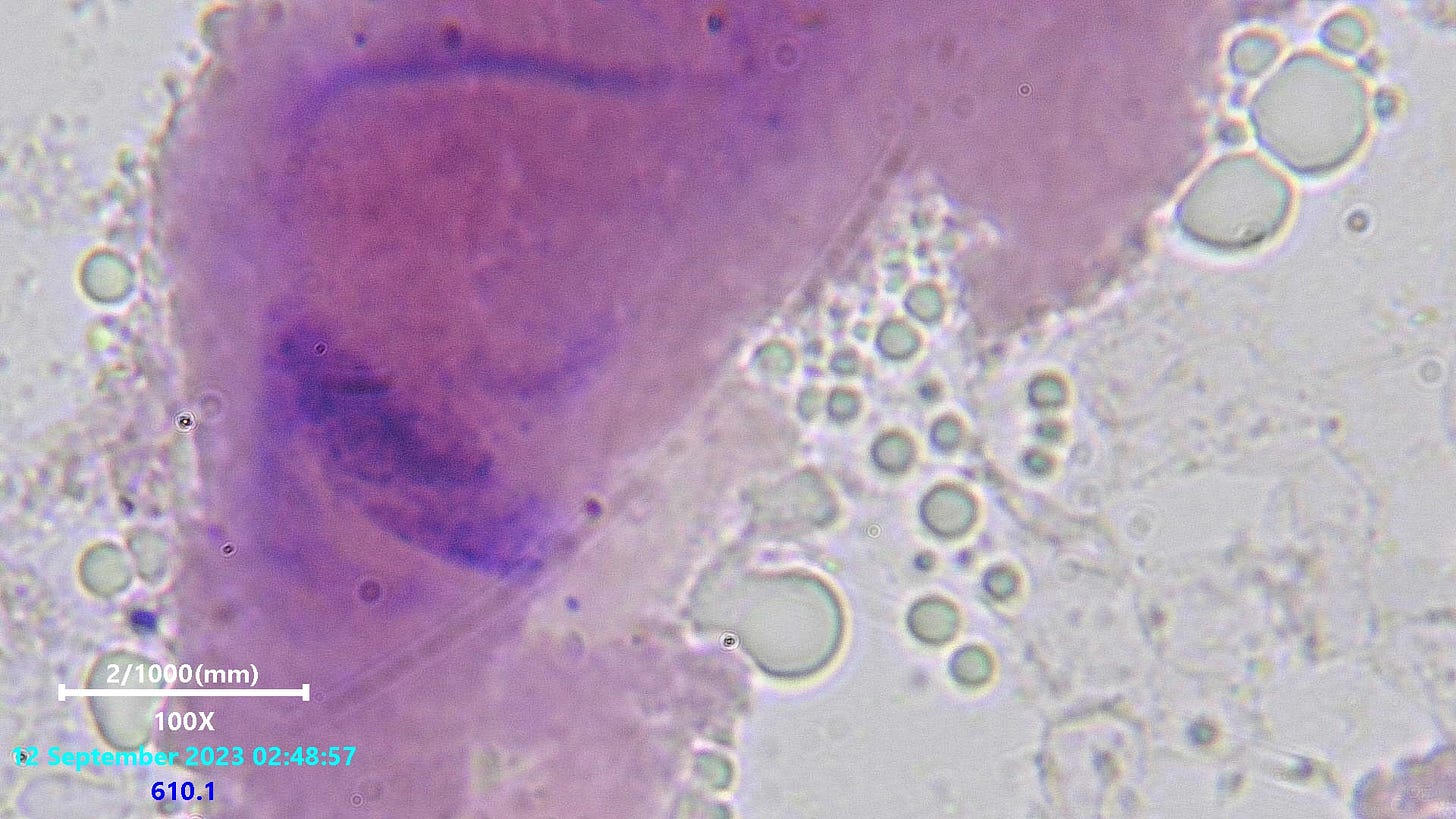
Taking a look at blood using gram stain technique that doesn’t look so bad in dark field reveals something very wrong indeed. The gels in the blood appear to of stained mostly negative gram, with some differing positive gram inclusions. Videos and other pictures below will highlight the likely possibility that the lipid droplets are slime. Mold type organisms. Possibly Dictyostelium, as suggested a long time ago as a possibility to someone on Researchgate who also saw this and could not easily sequence DNA from it. See the worrying behavior below which shows the entire slide quickly becomes swallowed by the surging fungus or amoeba.
The fungus like mass seems to join with the likewise appearing material forming from all the gels which stained negative gram. The bond is instant and clearly one and the same. Small cyst like bodies seem to be moving by the gel structures and also look to be of the same material
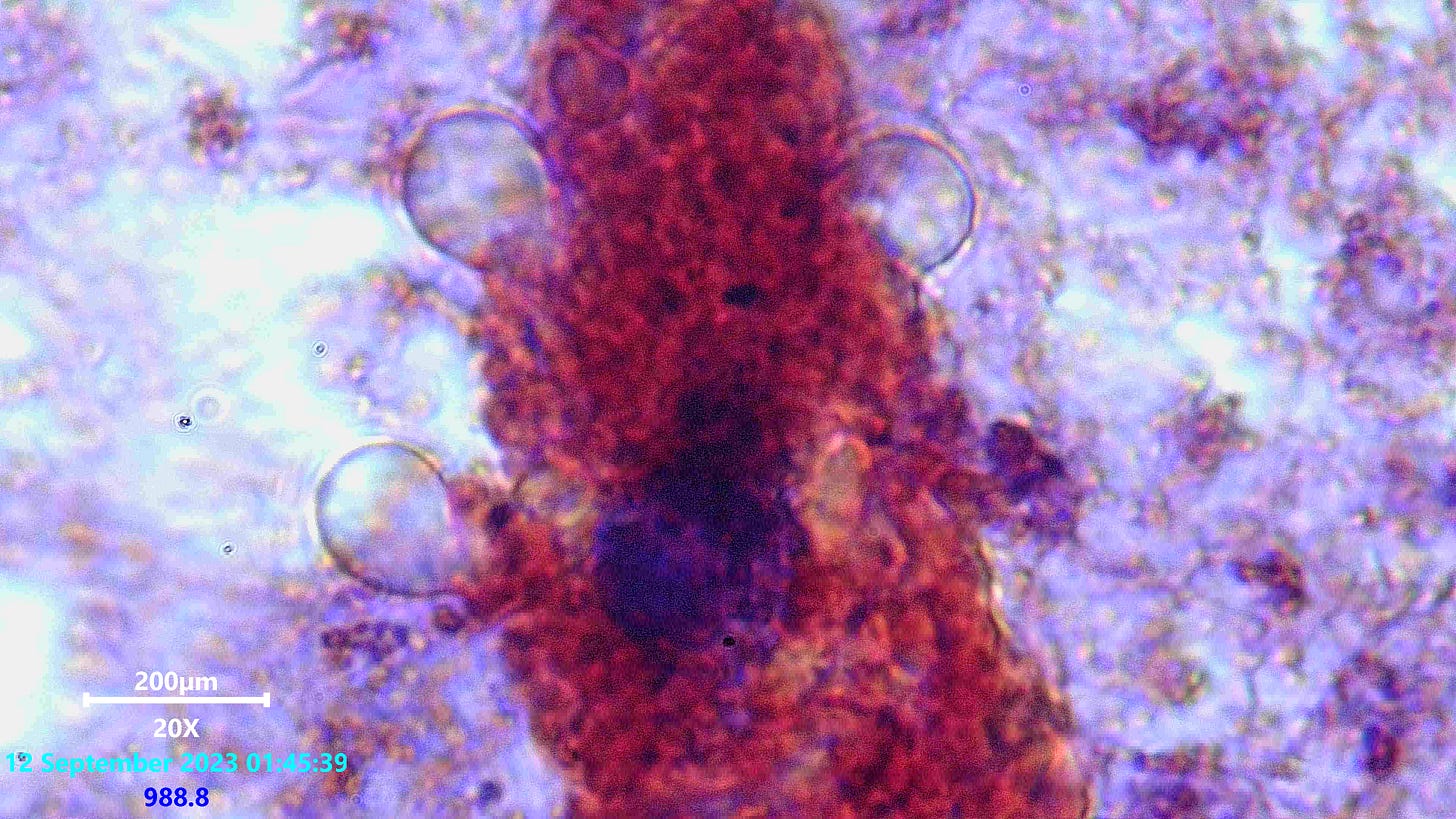
Some structures seemed to stain almost more red. I will have to go back and study into the possible colour variations later on. A positive gram mass can be seen centered
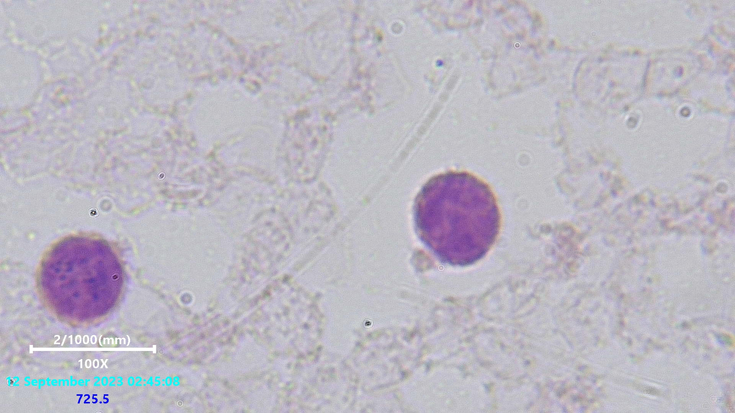
Bare with the images, there are a few variances. This one maybe contains a very small positive gram bacteria within. There were small positive gram organisms like these attached to many RBC’s. further below
Positive gram bacteria possibly, attached to RBC’
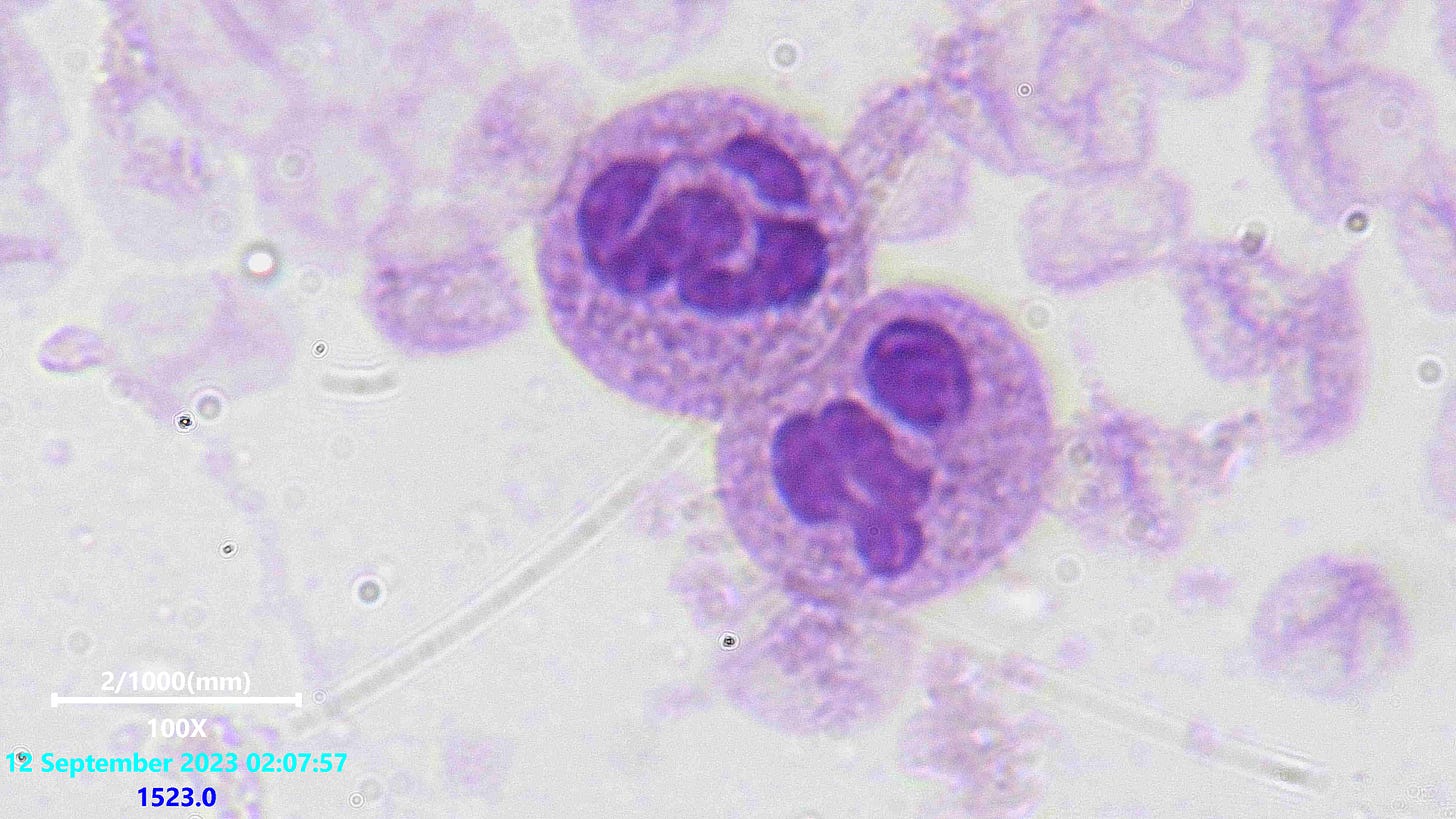
There were many white cells with fragmented nuclei or positive gram bacteria present such as this one on the left hand side above
Some slightly more regular looking ones here, thank god, i think
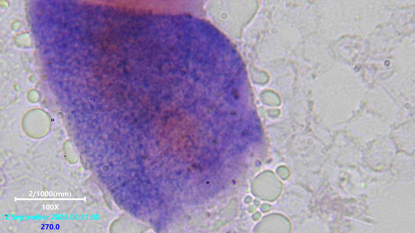
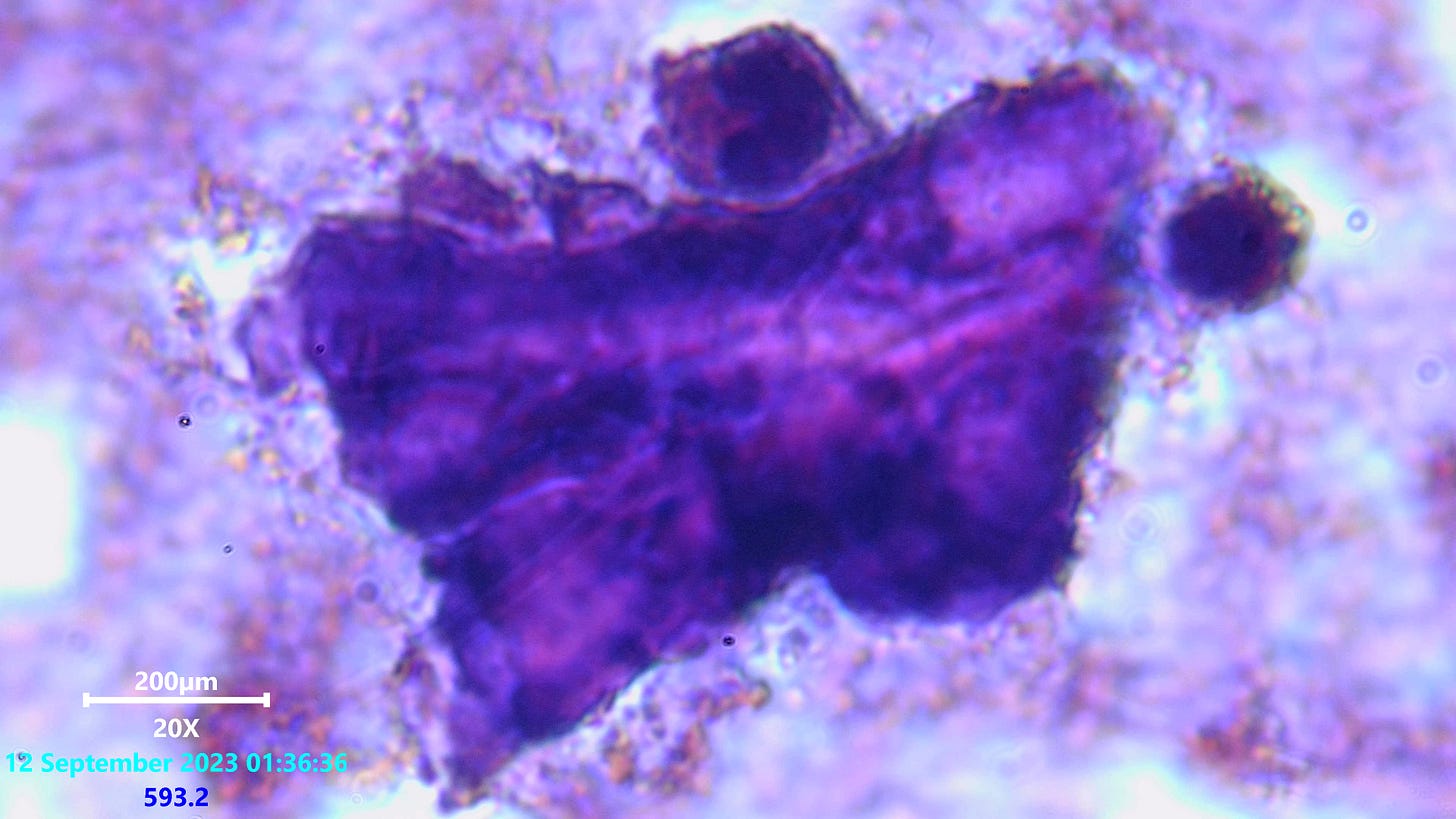
Large polymer like gel structures which stained quite differently but may also have been the victims of uneven staining in the procedure
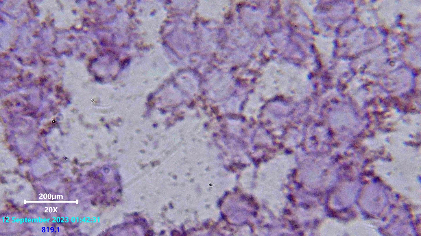
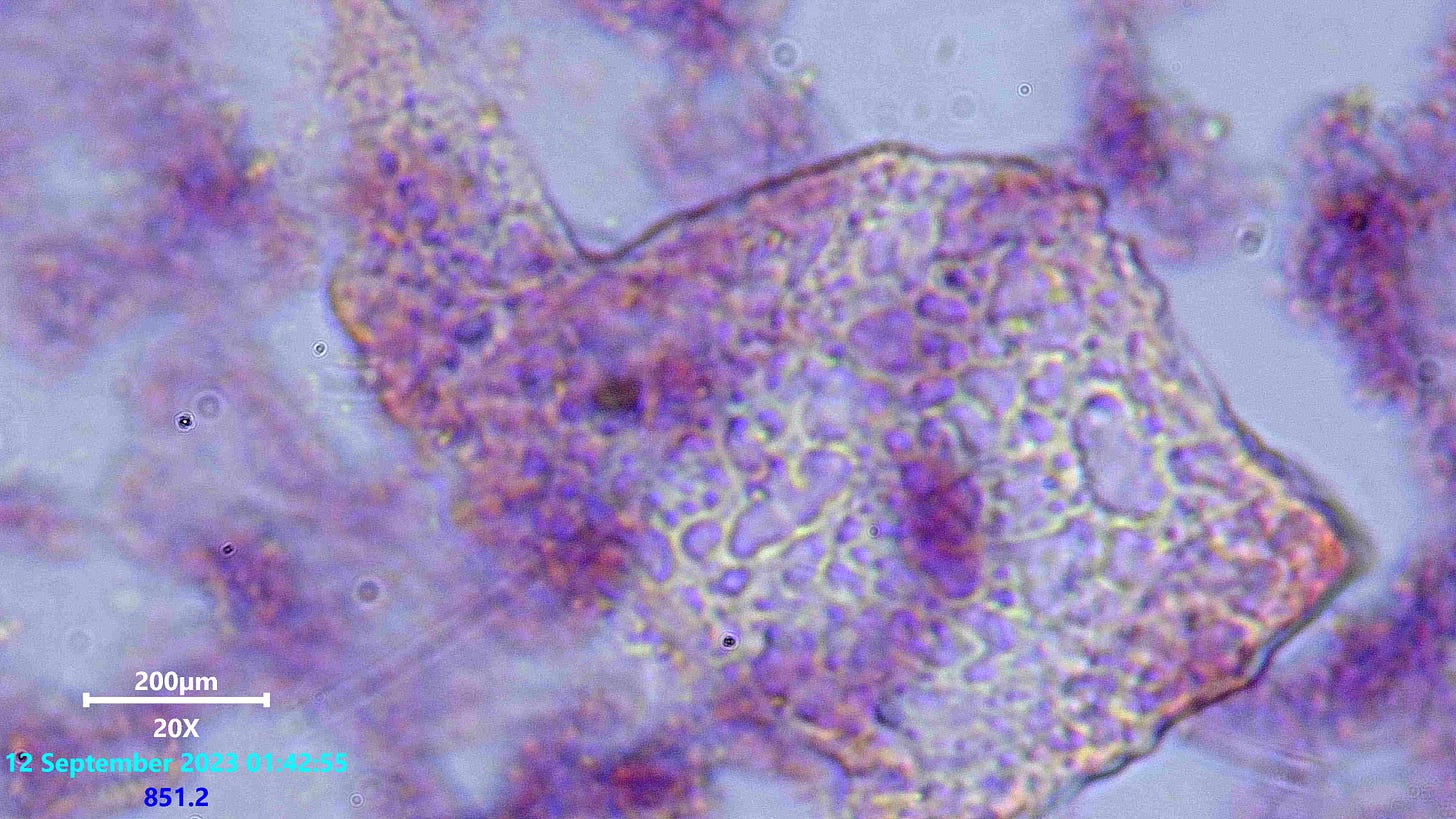
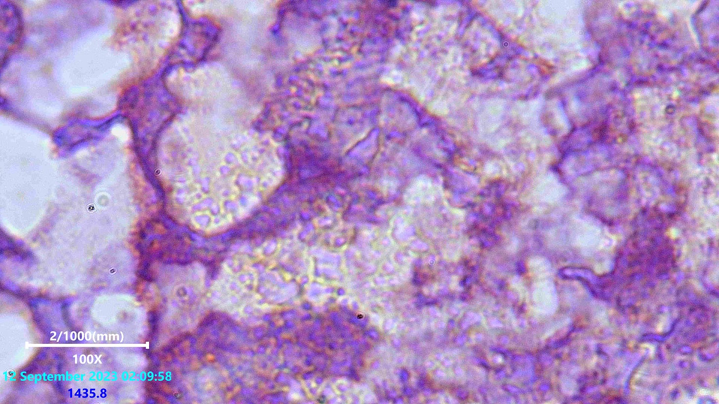
A clear indicator of possible fungus or slime mold type organisms again. Take a look at the plasmodium network like appearance, this appears like the the goopy bubbles seen coming from the gel structures. It can be seen morphing at times
And here again we can see the same plasmodial like network structures which appeared very quickly after preparing the slide. Some more investigation needs to be done here. But the only evidence of this stuff i find in dark field viewing of this blood looks more like the general issues we are all investigating now, assumed to be from shedding and vaxksines.
I will come back to this, it takes a long time to prep and view these stained slides. I find this very concerning. I have noticed this kind of activity going on now since i first got ill nearly 3 years ago. It seemed as if a few fibers were trying to form in this sample, although i focused mainly on other less known abnormalities here. This could be seperate to the main issue we generally refer to these days, but i have a feeling it is not and that this somehow fits between the nano, and Clifford Carnicom’s work.
Above is 100x (not 20x, sorry). See our bouncing friends from dark field look different in gels at 100x oil when stained.
Please do help support my joint efforts with others to find answers to the worlds biggest puzzle ever.
I am using KO-FI right now and appreciate you clicking the link to go there and contribute.
New lab gear is needed ;D......Thank You All !!!!!!


Has anyone tried oregano oil?
Fungus are easily treated with antibiotics…. Worth trying!