Self-assembly, Multi-lamellar vesicles and structures formed within the blood.
An update to material forming in the blood and forming in pharmaceuticals.
READ CAREFULLY, serves to provide evidence for self-assembly in brief detail.
Informed consent = uninformed trickery and complex, biologically altering, mass global experimentation.
For a very long time we have been studying many different foreign materials in the blood. It was maybe 5 months ago or more when we realized these thick membraned structures that were creating foreign material were possibly identifiable as a Multi-lamellar structure, before this we termed them constructor ring bubbles knowing they were advanced biosystems of some kind and indeed not bubbles. They have become a major component of our blood now and then clearly can be seen doing exactly what they look like they are doing………. producing material, self-assembly ! Unfortunately as we have been explaining for over a year now, it can be found in tap water and many other contaminated places. It is self replicating.
below is a bright field video taken of a stained swab sample under coverslip from last year 2023. What is happening in previous work is now becoming much more clear. The multiple dyes below show a transition of material chemistry forming up and joining other lipid structures, as they do this we can see reddish lipids forming with a thicker and more complex membrane membrane. This is the birth of another creative stage. This red product is likely the multilamellar structure being formed with hydrogel inside, just as we are seeing in blood.
Above video is of stained swabs recorded back at 10x speed.
Various gel like and lipid like material is laying out and forming in these samples. This is what devious self-assembly would look like, this is exactly the kind of process you would expect to see.
Above video is of stained swabs recorded back at 10x speed, this material may differ from the processes seen in the first video. The Dye used here was different, safranin. This can associate with same or differing material products depending on the dye properties. More observations are being made to interpret more specifics one these findings from last year.
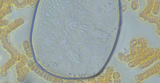
Above we see many unusual things going on. A large thick membraned structure with green overcast where chemical activities are occurring. Lipid structures are forming inside and in this case appear as snotty looking yellow structures. Long fibrin like multi-coloured crystals are forming from the membrane edge and may actually be a similar material to the snotty looking liposome membranes appearing. Internal production of synthetic material can be seen forming in the RBC’s surrounding the out membrane and also in the inner membrane centre of the large membrane. liposomes and other material can be seen between blood cells on the lower side of the image.
Similar structures can be seen here again in the image above. This time we see less colouration from chemical reactions inside these structures and more lipid structures identical to those which we see forming in vaccines, anaesthetics and on swabs or COVID-19 rapid antigen tests.

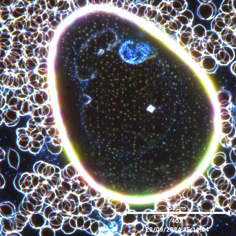
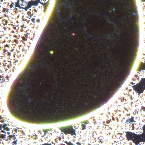
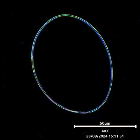
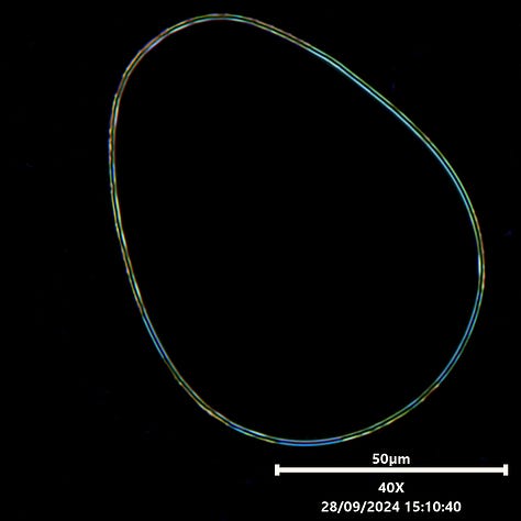
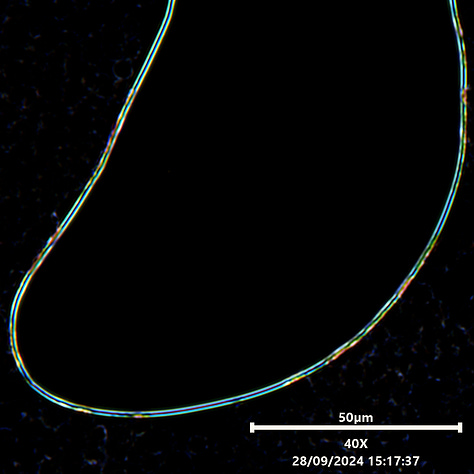
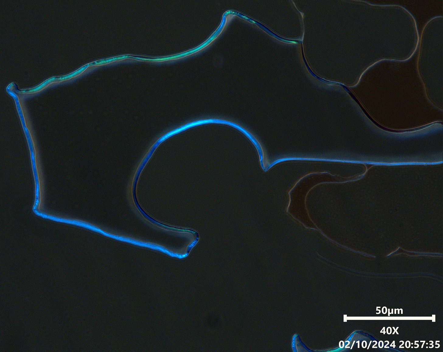
These images when enlarged show a view of multi-lamellar structures in normal light and in low light. The low light enables us to see complications in the membrane. The thickness is of note, the colouring variation in segments of the membrane, and movements of material in the membrane. Also worth noting is the luminous colours we often now see on these membranes in higher light as the top 3 images show and the same or similar colourful activity now being seen around and inside many RBC’s (red blood cells). not pictured here. New information has become apparent regarding lamellar hydrogels. These are known to have most of these properties. A thick fluorescent membrane, and gel like behaviour is described. Further more the papers talk about iridescence as a strong feature of materials and activates within. This would explain the confusion many had in deciding whether these structures were to be labelled a hydrogel, or a multilamellar vesicle. The behaviour and visual indications lend favour to the use of lamellar hydrogel. This kind of hydrogel is noted as being the most diverse and complex material that science has yet seen in terms of complex self assembly and customization.
The above shows a jig that was a little annoying to make. It is version 2 of the RGBW side lighter as referenced in science papers as a method of causing particles to fluoresce more brightly for viewing in dark field. The first was a success, this one could have been better and overall I was not satisfied with its function. Particles did not become enhanced, however membranes were very interesting as we see below, they captured this light angle well.
In both of these blood sample images (above and below) we find yet more indications of membrane complexity. It appears very polymer like and the very coloured green structures in these membranes showed plenty of complex movements or changes, particularly when the membrane would expand or shrink. We have many images using different techniques.
LASERS:
When lasers are used on samples, particularly UV lasers, We can enhanced the formation of some material and lipids can be seen expanding rapidly in most cases. The lasers transfer heat and also energy in the form of photons. Energy is required for most of these materials to continuing forming. Thermal activity is increased due to the energy of the laser, and light photons can also induce other electrical properties. Green laser has again shown no positive effects where I have tried or used them for imaging over the last few years. Green lasers had similar or same affects unfortunately, however it wasn’t a cure experiment, it was a detail imaging experiment that showed these affects occurring. Products are formed and the process sometimes collapses the membrane afterwards. Given the fact the those structures leave material behind lends me to understand that lasers speed up the processes but did not slow them or destroy them in any way whatsoever at any point in time. This is why none can provide good quality images of clean blood after using a green laser despite it being touted. Another debunk is shown in effect.
Above image: Multilamellar vesicles being formed up rapidly in blood samples. This are formed by smaller self assembling processes that cannot be seen as easily. The UV laser exposes moving waves around areas where the bright, thick membranes are expanding and constructing. This exploits activity in those regions quite well. The video is played at 10x speed using a 25x Fluotar objective.
In the image of my blood below, we can see again from a different perspective complex chemical processes occurring in the lamellar structures. The colour range is disturbing as you will see in following images. Synthetic liposomes are seen in varying formations. These appear as worm like structures, chained beads and follow no explainable similarities to parasites or their behaviour. Most of the foreign material that forms in our blood has a range of key identifiers that link all this material to association, not just by morphological observation. If you are familiar with the biowarfare that is Lyme disease and Morgellons then you may realize that there is uncanny significance with these features now seen in our blood. We are aware of these forms being referred to as liposomes, synthetic cell structures, and viruses in some literature and peer review papers. We have the full story on that and will be presenting it with the papers to compare for your self. I now believe I know what, how, and why I managed to treat my Lyme and MD so successfully, if still symptomatic at times. I am functioning fairly well if struggling a bit with my physical heath still. One of the key features that draws my attention is the connection between some tetracyclines, hydroxychloroquine, herbs and spice such as artemisia, turmeric, curcumin, spinach, garlic, and much more. All of these have bioactive compounds with one thing in common, and that seems to be relevant with the current pandemic version of this material we have been distributed. Please don’t twist my arm on that one, my detailed explanation will come with all the info. I have shared this with others in our research circle so I am never the only one holding information alone. It may not be that hugely significant, but we think it is something huge and may help us all going forward.
It is wise to remind the reader that these samples above and below are my blood samples as an unvaccinated 5 times swabbed champion of nothing. All blood looks vaccinated today. The extent of contamination presented here must be viewed with appropriate optical equipment and high end imagers to see the full presence of this devastating and altering material. Lower end microscopes miss huge details, colours, and unique features making everything look rather plain. Electron microscopy will serve little use in determining anything more than dark field in my opinion since all colour and clues are removed from the equation resulting in everyone looking at a grey, likely altered form of the material that bares no useful information. I believe high-end modified dark field microscopy, HPLC or mass gas spectrometry, and DNA sequencing (Not DNA quantification) alongside a few other helpful methods shall be key to unravelling the whole story with usable data to help ma the coded intention and functioning of this products sufficiently.
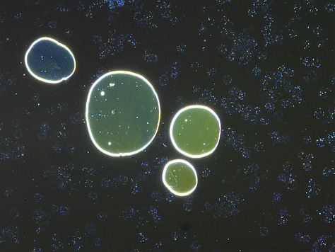


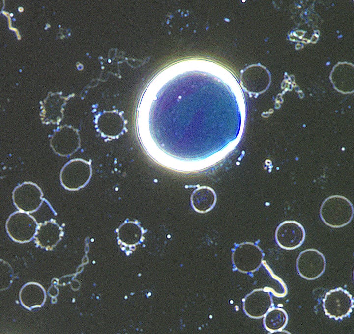
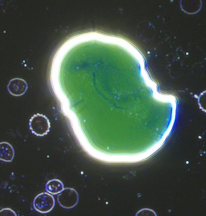


I ask kindly and without ego or greed that people reading this cite the source of these detailed discoveries. The reason being is we need support to keep going or to accelerate our work. That funding then goes to someone else who is just copying the painful and long work that others have done. There is a lot to add here and in other site sources, but the continuation of this work depends on support of others. We are aware that others will read these posts and quote the findings as their own work or discovery and then miss describe what we have shown by not understanding fully in order to direct funding on top of their daytime professions, I have been doing this every day and night for nearly 3 years or more and it is all I live for. I WANT TO FEEL 100% WELL AGAIN ONE DAY, AND I WANT EVERYONE ELSE TO HAVE THOSE ANSWERS TOO, come hell or high water!
Tip: if you see people claiming discovery of these materials and using the same words, check the article dates against ours ;). If someone else gives you tangible info and they are the source of that work or give citation and credit to the researchers then do consider supporting them too. Free journalism offered by small community news outlets is showing to be the only way we can get any tangible news out today, thanks to corrupt main stream media. Support the community voice’s out there and support our own freedoms.
The above sample is from Citonest ANAESTHEIC. It is the same stuff visually as what is seen in all blood now. Remember where or who this foreign material has come from and how it is now wide spread globally. A global agenda is in full play!
As always, please share and to help us get further towards affording an HPLC (liquid chromatography) machine. Got a lot of useful tools going so far but we can do much better at determining what this stuff is specifically doing. Thanks for reading !





Thank you, Karl. I'm praying for your well-being. That you are able to safely neutralize and or rid your body of these foreign entities and that you will be restored to full and vibrant HEALTH. In God's name and All that is Good. Amen
OooooHHHHH !!! WOW Karl, these images are probably the clearest most stunning and telling yet !
How on this earth can anyone in their right mind NOT see this damage ??
The TRUTH will always win OUT,
God bless you man against the microbes - and - damn the scum behind all this..... KK