


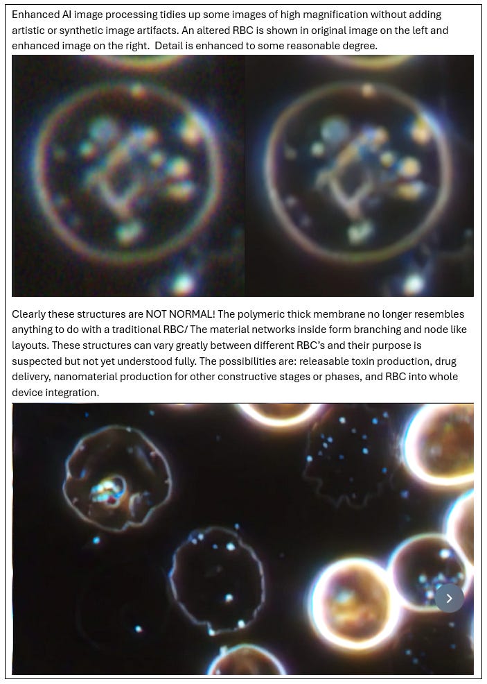
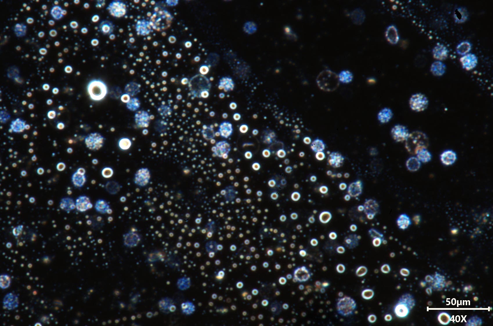
Synthetically altered blood @ 30,000 lumens, Darkfield. ---> LOOK <---
Blood cell images from the latest darkfield setup. Stunning detail !

Are there really references that tell us how birefringence and iridescence should behave in structures such as natural blood cells, Synthetic blood cells, liposomes, vesicles, exosomes, and other artificial cell types? Yes…there is.

This paper from the ‘Journal of Biomedical Optics’ shows measurement and characterization of optical phenomenon known to natural blood cells. In this case linearly polarized green light (λ ¼ 546 nm) is used to measure these optical features. However, nothing in this paper is even close to being able to plot, predict, or explain what is seen in my work results.

Much of the colourful effect seen in my images could only possibly be occurring more so at the curved edges of the blood cell and in curved areas near the concave dip. As this paper shows, intensely fluorescing and transforming shapes withing the cell are not part of optical effects. Instead, it was it is to be expected of complex vesicles with multiple membranes forming internally. Below is an extract from my private journal. Everyone’s blood can be seen forming this processes and much more clearly and cleanly after time on the slide has passed.

Focus into the geometry and optical specifics is still being profiled, but at this stage all massive arrows and current science knowledge direct us only to bio-engineering. The purpose of endless profiling like we can see in my other extract below is more to give us extra information regarding what type of synthetic process this is and how it all works, not to decide if the material is natural or synthetic since we can see that it is synthetic already for reasons above and more.
So, as we can see there are many different methods of characterizing optical and material data from samples, even just using software based scientific tools. morphology tools, FFT analysis, spacing, sizing, and exact geometries are all being catalogued for a larger perspective and explanation regarding the variances and behaviors of this synthetic material and its processes. I do not advise using LLM/AI to output graphs and charts for reliable data. These clever platforms do not set optimal parameters in the image processing chain and this leads to it outputting vaguely useful data, but not accurate data. I used chatGPT’s ability to take an image, then perform certain algorithm based analysis and compared it to manual parameter entry or tweaked script/macro output in applications such as ImageJ and other scientific processing apps. The results were what I would say was close, but definitely not close enough for science.
An example of FFT analysis performed on human hair from my personal journal can be seen below. The LLM/AI does not provide clear and well organized frequency based visual analysis of structures in question. Manual calibration of parameter settings truly gives optimized processing like we see in my page below.

Typically different structure FFT analysis using LLM/AI provided output which was far less distinct, of lower resolution, and with many spacial errors. See the FFT graph below for an example, background artifacts were strong in the outer detail areas.

Without further deviation into technical methods being employed for optical analysis we can see how important this information is. It is a high resolution mapping of many optical properties that can indicate material properties and construction pathways to some reasonable extent when correlated and compared to relevant databases for which I have been collecting from online. Everything is useful in presenting the full picture. What this all means will take more time in terms of interpreting the data, but some jobs have already been compared with interesting implications for material complexity and types.
If you appreciate my constant efforts and time please help the cause and speed up research by contributing to equipment supplies and more. This cannot continue without the help other other living men and women !

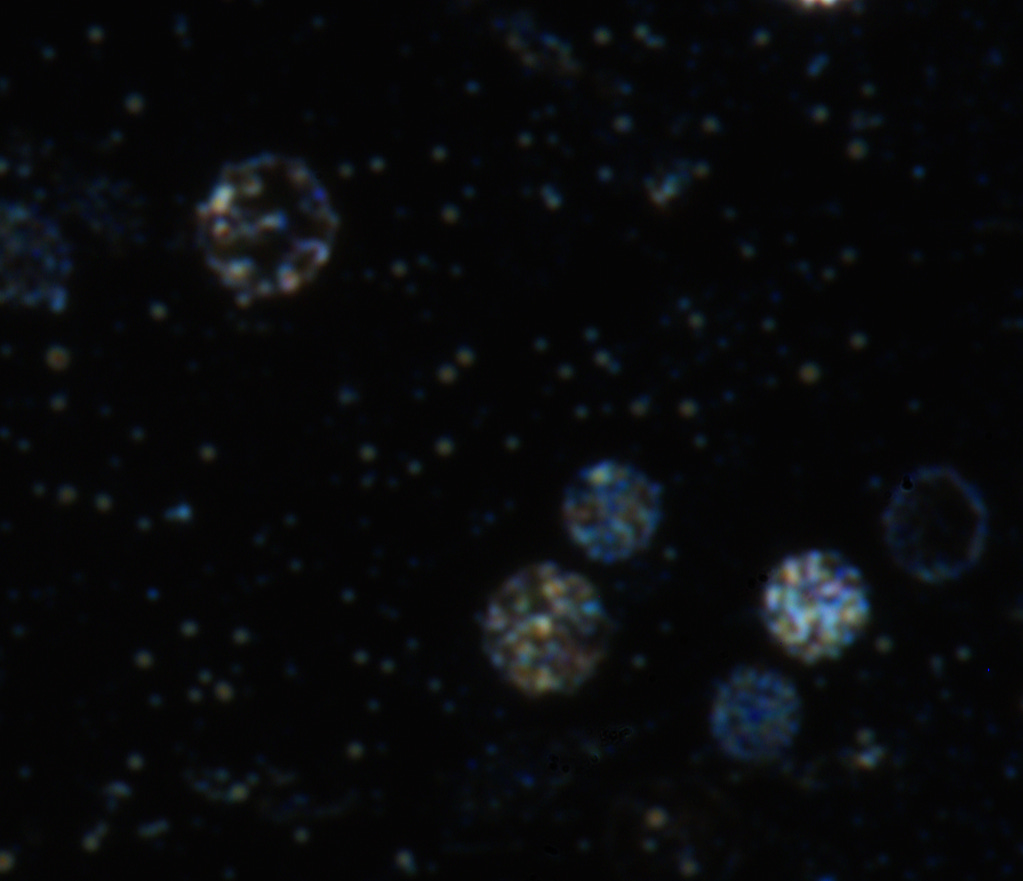
Above: recent collage of images taken from unvaccinated blood. At this stage trying to guess what exact unique structures are forming in terms of assembled material is still a fools erund. What we can confirm from this quality of imagery is that we are looking at constructs only known to the field of advanced nanotechnology. It isn’t often in biological science you can say much about something you cannot see and yet still likely be right. But what is in the images is far too obvious to confuse with the appearance of natural biological structures. Previous science in biology does not align with what we see here, but synthetic sciences do without any debate. This is a plausible conclusion given previous data. In this one collage alone we can tell several key factors which are synthetic mostly and only features. Membrane thickness of RBC’s, Inner layer vesicle membranes, varying membrane optical appearance, Polymeric type membrane attributes, Internal and observable morphological processes leading up-to large complex structures of programmed nature, Presence of Hybrid lipid nanoparticles, extreme iridescence, and multi-domain inner vesicles showing separate material properties. This isn’t behavior of natural biology, its well known attributes applied to synthetic cell vesicles and altered cellular biology.
So, no ignorant nurse or doctor can really look at these while pretending they know it is normal, because it isn’t and they most often have very limited experience in looking at blood themselves, or have never been allowed to use a dark field microscope since western medicine pretty much forbids it. They also have absolutely no experience or understanding in the field of nanotechnology/synthetic biological sciences. Thank god we have some awake and open minded doctors still thinking, it isn’t many sadly, but its something. It seems we have too many of us which feel okay about fact checking things we haven’t even self investigated or have any background in. This doesn’t help public awareness in a corrupt society and agenda based global takeover scenario either, we need to stand together! The end result of all this isn’t meant to resort to you being happy and having your freedoms as Klauss Schwabb explained at the WEF. Its about AI, connected everything, dominance, population control, and future slavery for your loved ones and friends. I think many have started to realize that researchers, doctors, and some scientists were not making this stuff up the last 4 or 5 years. I only wonder what some people think is going to happen if they just do nothing? Many of us think we know, but we didn’t guess it all, the answers were publicly provided in full knowing the public would not take them seriously. People just didn’t listen and made excuses for why not to look, or why it must of all just been fake noise they’ve been hearing.
Since we’ve made some headway on some of the projects been happening lately I shall leave you with a whole bunch of new images to feast the eye balls on. I dont want to say too much now since I have more to do, obviously. I am really pleased with the microscope upgrades and performance combined with software analysis techniques so far. It is best to only use high-resolution, quality work as accurate images processed into logical data and this setup certainly excels there so far. I am also excited to see where these libraries of data will take things. Other analysis is still running alongside also.
Back to cells from hell !
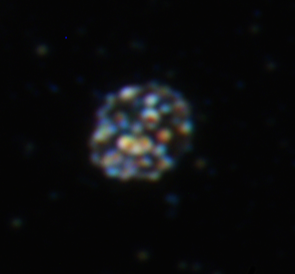
This cell is enlarged with objective aperture mostly closed, It enhances the ability to differentiate colour inside the cell and reveals properties of material which is different, ordered, and complex. What it loses is resolution and some light power, but it gives priceless insight in its own right. These internals are not known to normal human biology either. These have spread throughout the population mostly since the pandemic period. They may have been seen in older man-made and controversial diseases, but that is another story.

Different altered cell sizes again, altered membranes, and almost what looks like blue structures possibly acting as linkers in this stage of the synthetic framework (a sort of joiner Lego brick).


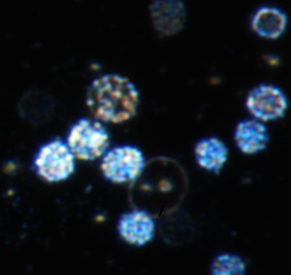
It is amazing, yet disturbing to see such a beautiful exhibition of all these synthetic details. At the same time as feeling privileged and please the microscope is performing so well, I am mortified. These variances and distinctly bold alteration effects are so easy to follow and capture now.

It would seem the blue cells are different to the clear, larger cells which have generally quite different material composition inside. This is a sign of multiple processes and extreme product complexity in this view alone.

In these post we do not focus too heavily on other structures but we can see some lipid trails exhibiting notable optical presentation which seems closer to that of anything but natural lipids when comparing other studies.

Upstairs here we have another really detailed shot inside this RBC top left. Make no mistake, that isn’t what proteins stripping or cell degradation looks like. Yet again, for many reasons including, size, difference, morphology, and layout this cannot be confused for natural biological material processes. These materials are polymerically chained often, have ordered or programmed assembly patterns, they are large materials after construction. ranging from low nm size all the way up to 2 um and more if assembling chains or other composite structures.
NOTHING TO SEE HERE………BACK AWAY FROM THE BLOOD VEHICLE!


A REMINDER TO OTHER RESEARCHERS AND SCIENTISTS!
High-end optical equipment, powerful lighting, high-quality objectives with adjustable aperture, and industrial grade imagers are required to see this much detail and realism. I hope others explore the same, the right people really need to of course. I am reluctant to give away my personal system modification and designs freely. I am sick of everyone claiming things for themselves and not returning the favor in a productive, helpful, or meaningful way. Ill just sit on it for now, unless something comes up. I have little interest in personal gain since i don’t live a normal life with all this going on anyway. But, this is not a cheap line of work either and progress comes far quicker with resources as always.

Almost out of the vesicle form inside the above RBC and headed towards the material fabrication stages.
above and below shows areas of dense and slowly forming artificial lipid structures, some reaching the large and massive lamellar vesicle sizes. Another detail many will never see with many microscopes, the colourful and differing material properties.

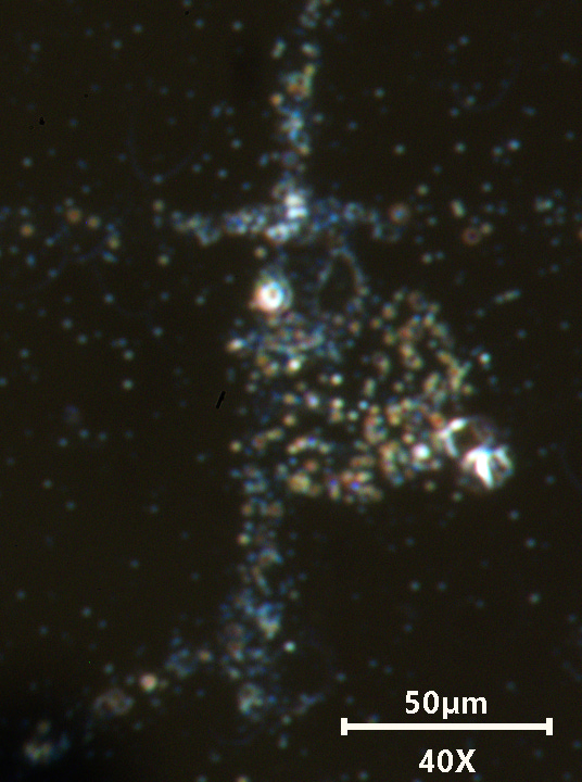
Networked materials with crystal phases appearing in some areas. (above).

The colours seen in this cell are almost fluorescent, the outer membrane in mid transition stage as it loses any last natural RBC features and begins to transform into a thicker polymeric vesicle type cell. Watching the various transitions can be very intriguing and there is no mistaking these repeat transitions processes.


Another display of colourful lipid-polymer networks floated up toward the slide cover slip. Various transhuman RBC’s in the plasma just below the lipids and left/right.


Our video below catches smaller liposomes with blue particles moving internally on the left next to the RBC which has similar membrane appearance and highly reflective material instead. Other cell stages and types also present. Spherical bodies seen floating in the plasma of various sizes and types. Nanoparticles, lipid structures, and other debris.
Thank you everyone! Those who have supported have really helped get this cutting edge viewing ability to form. A small amount of contribution over the 2 years has taken me leaps and bounds while working with what little is affordable to me. The Thermo nicolet FT-IR 6700 spectrometer still needs parts replaced or whole unit to be replaced since it isn’t functioning yet still. It has a minor issue, but it takes a long time to troubleshoot complex technology without service documents that pharma industries stop you having for the very reason of controlling public research and knowledge. You would think they like to encourage science, but that isn’t the case. They do the same with all types of high-end lab gear which could validate or detect chemical materials accurately, read small signals, and more, they hide technology and access to it behind contracts and pay walls. That isn’t an accident, it is controlling knowledge. Those issues can be tackled with planning and special arrangements. but, it still costs even on used cost basis with parts scavenging.
If you appreciate my constant efforts and time, please help the cause and speed up research by contributing to equipment supplies and more. This cannot continue without the help other other living men and women !
Thanks for supporting 🙏
Thank you Karl for all your dedication. I wish more people were awake to this but no one wants to hear it. It’s very frustrating.